6YQG

Crystal structure of native Phycocyanin in spacegroup P63 at 1.45 Angstroms.


Experimental Data Snapshot

  • Method: X-RAY DIFFRACTION
  • Resolution: 1.45 Å
  • R-Value Free: 0.208 
  • R-Value Work: 0.178 
  • R-Value Observed: 0.179 

wwPDB Validation   3D Report Full Report


Ligand Structure Quality Assessment 


This is version 1.2 of the entry. See complete history


Literature

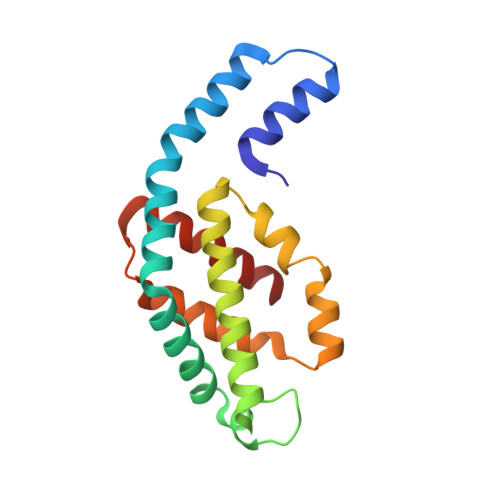
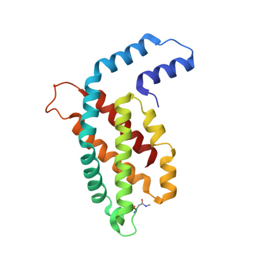
C-phycocyanin as a highly attractive model system in protein crystallography: unique crystallization properties and packing-diversity screening.

Sarrou, I.Feiler, C.G.Falke, S.Peard, N.Yefanov, O.Chapman, H.

(2021) Acta Crystallogr D Struct Biol 77: 224-236

  • DOI: https://doi.org/10.1107/S2059798320016071
  • Primary Citation of Related Structures:  
    6YPQ, 6YQ8, 6YQG, 6YYJ

  • PubMed Abstract: 

    The unique crystallization properties of the antenna protein C-phycocyanin (C-PC) from the thermophilic cyanobacterium Thermosynechococcus elongatus are reported and discussed. C-PC crystallizes in hundreds of significantly different conditions within a broad pH range and in the presence of a wide variety of precipitants and additives. Remarkably, the crystal dimensions vary from a few micrometres, as used in serial crystallography, to several hundred micrometres, with a very diverse crystal morphology. More than 100 unique single-crystal X-ray diffraction data sets were collected from randomly selected crystals and analysed. The addition of small-molecule additives revealed three new crystal packings of C-PC, which are discussed in detail. The high propensity of this protein to crystallize, combined with its natural blue colour and its fluorescence characteristics, make it an excellent candidate as a superior and highly adaptable model system in crystallography. C-PC can be used in technical and methods development approaches for X-ray and neutron diffraction techniques, and as a system for comprehending the fundamental principles of protein crystallography.


  • Organizational Affiliation

    Centre for Free-Electron Laser Science, DESY, Notkestrasse 85, 22607 Hamburg, Germany.


Macromolecules
Find similar proteins by:  (by identity cutoff)  |  3D Structure
Entity ID: 1
MoleculeChains Sequence LengthOrganismDetailsImage
C-phycocyanin alpha chain162Thermosynechococcus vestitusMutation(s): 0 
UniProt
Find proteins for P50032 (Thermosynechococcus vestitus (strain NIES-2133 / IAM M-273 / BP-1))
Explore P50032 
Go to UniProtKB:  P50032
Entity Groups  
Sequence Clusters30% Identity50% Identity70% Identity90% Identity95% Identity100% Identity
UniProt GroupP50032
Sequence Annotations
Expand
  • Reference Sequence
Find similar proteins by:  (by identity cutoff)  |  3D Structure
Entity ID: 2
MoleculeChains Sequence LengthOrganismDetailsImage
C-phycocyanin beta chain172Thermosynechococcus vestitusMutation(s): 0 
UniProt
Find proteins for P50033 (Thermosynechococcus vestitus (strain NIES-2133 / IAM M-273 / BP-1))
Explore P50033 
Go to UniProtKB:  P50033
Entity Groups  
Sequence Clusters30% Identity50% Identity70% Identity90% Identity95% Identity100% Identity
UniProt GroupP50033
Sequence Annotations
Expand
  • Reference Sequence
Experimental Data & Validation

Experimental Data

  • Method: X-RAY DIFFRACTION
  • Resolution: 1.45 Å
  • R-Value Free: 0.208 
  • R-Value Work: 0.178 
  • R-Value Observed: 0.179 
  • Space Group: P 63
Unit Cell:
Length ( Å )Angle ( ˚ )
a = 108.208α = 90
b = 108.208β = 90
c = 66.05γ = 120
Software Package:
Software NamePurpose
PHENIXrefinement
XDSdata reduction
Aimlessdata scaling
PHASERphasing

Structure Validation

View Full Validation Report


Ligand Structure Quality Assessment 


Entry History 

Deposition Data

Revision History  (Full details and data files)

  • Version 1.0: 2021-01-20
    Type: Initial release
  • Version 1.1: 2021-02-17
    Changes: Database references
  • Version 1.2: 2024-01-24
    Changes: Data collection, Database references, Refinement description