6ZN3

Plasmodium facliparum glideosome trimeric sub-complex


Experimental Data Snapshot

  • Method: X-RAY DIFFRACTION
  • Resolution: 2.51 Å
  • R-Value Free: 0.238 
  • R-Value Work: 0.200 
  • R-Value Observed: 0.202 

wwPDB Validation   3D Report Full Report


This is version 1.2 of the entry. See complete history


Literature

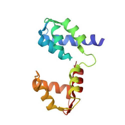
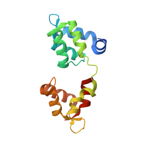
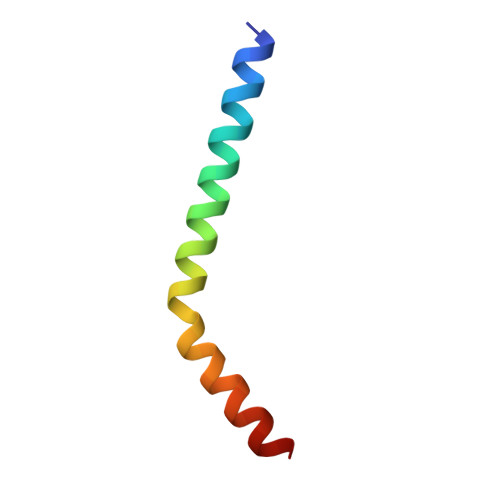
Structural role of essential light chains in the apicomplexan glideosome.

Pazicky, S.Dhamotharan, K.Kaszuba, K.Mertens, H.D.T.Gilberger, T.Svergun, D.Kosinski, J.Weininger, U.Low, C.

(2020) Commun Biol 3: 568-568

  • DOI: https://doi.org/10.1038/s42003-020-01283-8
  • Primary Citation of Related Structures:  
    6TJ3, 6TJ4, 6TJ5, 6TJ6, 6TJ7, 6ZN3

  • PubMed Abstract: 

    Gliding, a type of motility based on an actin-myosin motor, is specific to apicomplexan parasites. Myosin A binds two light chains which further interact with glideosome associated proteins and assemble into the glideosome. The role of individual glideosome proteins is unclear due to the lack of structures of larger glideosome assemblies. Here, we investigate the role of essential light chains (ELCs) in Toxoplasma gondii and Plasmodium falciparum and present their crystal structures as part of trimeric sub-complexes. We show that although ELCs bind a conserved MyoA sequence, P. falciparum ELC adopts a distinct structure in the free and MyoA-bound state. We suggest that ELCs enhance MyoA performance by inducing secondary structure in MyoA and thus stiffen its lever arm. Structural and biophysical analysis reveals that calcium binding has no influence on the structure of ELCs. Our work represents a further step towards understanding the mechanism of gliding in Apicomplexa.


  • Organizational Affiliation

    Centre for Structural Systems Biology (CSSB), Notkestrasse 85, D-22607, Hamburg, Germany.


Macromolecules
Find similar proteins by:  (by identity cutoff)  |  3D Structure
Entity ID: 1
MoleculeChains Sequence LengthOrganismDetailsImage
Myosin essential light chain ELC
A, D, G, J, M
135Plasmodium falciparum 3D7Mutation(s): 0 
Gene Names: PF3D7_1017500
UniProt
Find proteins for Q8IJM4 (Plasmodium falciparum (isolate 3D7))
Explore Q8IJM4 
Go to UniProtKB:  Q8IJM4
Entity Groups  
Sequence Clusters30% Identity50% Identity70% Identity90% Identity95% Identity100% Identity
UniProt GroupQ8IJM4
Sequence Annotations
Expand
  • Reference Sequence
Find similar proteins by:  (by identity cutoff)  |  3D Structure
Entity ID: 2
MoleculeChains Sequence LengthOrganismDetailsImage
Myosin A tail domain interacting protein
B, E, H, K, N
147Plasmodium falciparum 3D7Mutation(s): 0 
Gene Names: PF3D7_1246400
UniProt
Find proteins for Q8I4W8 (Plasmodium falciparum (isolate 3D7))
Explore Q8I4W8 
Go to UniProtKB:  Q8I4W8
Entity Groups  
Sequence Clusters30% Identity50% Identity70% Identity90% Identity95% Identity100% Identity
UniProt GroupQ8I4W8
Sequence Annotations
Expand
  • Reference Sequence
Find similar proteins by:  (by identity cutoff)  |  3D Structure
Entity ID: 3
MoleculeChains Sequence LengthOrganismDetailsImage
Myosin-A
C, F, I, L, O
43Plasmodium falciparum 3D7Mutation(s): 0 
Gene Names: PF13_0233
UniProt
Find proteins for Q8IDR3 (Plasmodium falciparum (isolate 3D7))
Explore Q8IDR3 
Go to UniProtKB:  Q8IDR3
Entity Groups  
Sequence Clusters30% Identity50% Identity70% Identity90% Identity95% Identity100% Identity
UniProt GroupQ8IDR3
Sequence Annotations
Expand
  • Reference Sequence
Experimental Data & Validation

Experimental Data

  • Method: X-RAY DIFFRACTION
  • Resolution: 2.51 Å
  • R-Value Free: 0.238 
  • R-Value Work: 0.200 
  • R-Value Observed: 0.202 
  • Space Group: P 43
Unit Cell:
Length ( Å )Angle ( ˚ )
a = 211.88α = 90
b = 211.88β = 90
c = 75.46γ = 90
Software Package:
Software NamePurpose
REFMACrefinement
PHENIXrefinement
STARANISOdata scaling
PHASERphasing
XDSdata reduction

Structure Validation

View Full Validation Report


Entry History & Funding Information

Deposition Data


Funding OrganizationLocationGrant Number
Swedish Research CouncilSweden621-2013-5905
Joachim Herz StiftungGermany800026

Revision History  (Full details and data files)

  • Version 1.0: 2020-10-21
    Type: Initial release
  • Version 1.1: 2020-10-28
    Changes: Database references
  • Version 1.2: 2024-01-31
    Changes: Data collection, Database references, Refinement description