RCSB PDB - 6W25: Crystal structure of the Melanocortin-4 Receptor (MC4R) in complex with SHU9119

 6W25

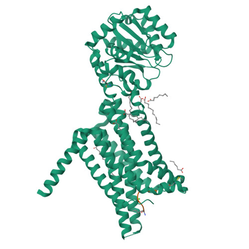
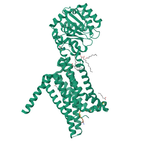
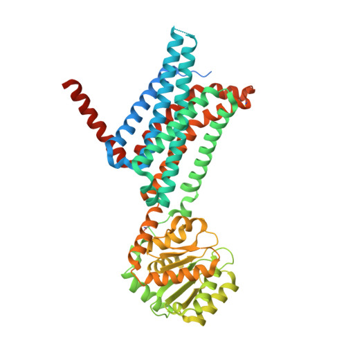
Crystal structure of the Melanocortin-4 Receptor (MC4R) in complex with SHU9119


Experimental Data Snapshot

  • Method: X-RAY DIFFRACTION
  • Resolution: 2.75 Å
  • R-Value Free: 0.259 
  • R-Value Work: 0.233 
  • R-Value Observed: 0.234 

Starting Model: experimental
View more details

wwPDB Validation   3D Report Full Report


Ligand Structure Quality Assessment 

Created with Raphaël 2.3.0Worse 01 BetterLigand structure goodness of fit to experimental dataBest fitted OLAClick on this verticalbar to view details

This is version 2.2 of the entry. See complete history


Literature

Determination of the melanocortin-4 receptor structure identifies Ca2+as a cofactor for ligand binding.

Yu, J.Gimenez, L.E.Hernandez, C.C.Wu, Y.Wein, A.H.Han, G.W.McClary, K.Mittal, S.R.Burdsall, K.Stauch, B.Wu, L.Stevens, S.N.Peisley, A.Williams, S.Y.Chen, V.Millhauser, G.L.Zhao, S.Cone, R.D.Stevens, R.C.

(2020) Science 368: 428-433

  • DOI: https://doi.org/10.1126/science.aaz8995
  • Primary Citation of Related Structures:  
    6W25

  • PubMed Abstract: 

    The melanocortin-4 receptor (MC4R) is involved in energy homeostasis and is an important drug target for syndromic obesity. We report the structure of the antagonist SHU9119-bound human MC4R at 2.8-angstrom resolution. Ca 2+ is identified as a cofactor that is complexed with residues from both the receptor and peptide ligand. Extracellular Ca 2+ increases the affinity and potency of the endogenous agonist α-melanocyte-stimulating hormone at the MC4R by 37- and 600-fold, respectively. The ability of the MC4R crystallized construct to couple to ion channel Kir7.1, while lacking cyclic adenosine monophosphate stimulation, highlights a heterotrimeric GTP-binding protein (G protein)-independent mechanism for this signaling modality. MC4R is revealed as a structurally divergent G protein-coupled receptor (GPCR), with more similarity to lipidic GPCRs than to the homologous peptidic GPCRs.


  • Organizational Affiliation

    iHuman Institute, ShanghaiTech University, Pudong, Shanghai 201210, China.


Macromolecules
Find similar proteins by:  (by identity cutoff)  |  3D Structure
Entity ID: 1
MoleculeChains Sequence LengthOrganismDetailsImage
Melanocortin receptor 4,GlgA glycogen synthase,Melanocortin receptor 4535Homo sapiensPyrococcus abyssi GE5Mutation(s): 5 
Gene Names: MC4RPAB2292
Membrane Entity: Yes 
UniProt & NIH Common Fund Data Resources
Find proteins for Q9V2J8 (Pyrococcus abyssi (strain GE5 / Orsay))
Explore Q9V2J8 
Go to UniProtKB:  Q9V2J8
Find proteins for P32245 (Homo sapiens)
Explore P32245 
Go to UniProtKB:  P32245
PHAROS:  P32245
GTEx:  ENSG00000166603 
Entity Groups  
Sequence Clusters30% Identity50% Identity70% Identity90% Identity95% Identity100% Identity
UniProt GroupsQ9V2J8P32245
Sequence Annotations
Expand
  • Reference Sequence

Find similar proteins by:  Sequence   |   3D Structure  

Entity ID: 2
MoleculeChains Sequence LengthOrganismDetailsImage
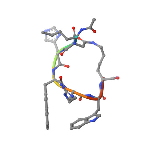
SHU91199synthetic constructMutation(s): 0 
Sequence Annotations
Expand
  • Reference Sequence
Small Molecules
Ligands 2 Unique
IDChains Name / Formula / InChI Key2D Diagram3D Interactions
OLA
Query on OLA

Download Ideal Coordinates CCD File 
D [auth A]
E [auth A]
F [auth A]
G [auth A]
H [auth A]
OLEIC ACID
C18 H34 O2
ZQPPMHVWECSIRJ-KTKRTIGZSA-N
CA
Query on CA

Download Ideal Coordinates CCD File 
C [auth A]CALCIUM ION
Ca
BHPQYMZQTOCNFJ-UHFFFAOYSA-N
Modified Residues  2 Unique
IDChains TypeFormula2D DiagramParent
YCM
Query on YCM
A
L-PEPTIDE LINKINGC5 H10 N2 O3 SCYS
NLE
Query on NLE
B
L-PEPTIDE LINKINGC6 H13 N O2LEU
Experimental Data & Validation

Experimental Data

  • Method: X-RAY DIFFRACTION
  • Resolution: 2.75 Å
  • R-Value Free: 0.259 
  • R-Value Work: 0.233 
  • R-Value Observed: 0.234 
  • Space Group: C 1 2 1
Unit Cell:
Length ( Å )Angle ( ˚ )
a = 163.91α = 90
b = 44.49β = 97.47
c = 88.05γ = 90
Software Package:
Software NamePurpose
PHASERphasing
BUSTERrefinement
PDB_EXTRACTdata extraction
XDSdata reduction
XDSdata scaling

Structure Validation

View Full Validation Report


Ligand Structure Quality Assessment 

Created with Raphaël 2.3.0Worse 01 BetterLigand structure goodness of fit to experimental dataBest fitted OLAClick on this verticalbar to view details

Entry History 

Revision History  (Full details and data files)

  • Version 1.0: 2020-04-29
    Type: Initial release
  • Version 1.1: 2020-05-06
    Changes: Database references, Refinement description
  • Version 2.0: 2020-09-09
    Changes: Advisory, Atomic model, Database references, Derived calculations, Structure summary
  • Version 2.1: 2023-10-18
    Changes: Data collection, Database references, Refinement description
  • Version 2.2: 2023-11-15
    Changes: Data collection, Derived calculations