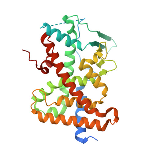
7AXI

Crystal structure of the hPXR-LBD in complex with estradiol and cis-chlordane


Experimental Data Snapshot

  • Method: X-RAY DIFFRACTION
  • Resolution: 2.15 Å
  • R-Value Free: 0.203 
  • R-Value Work: 0.173 
  • R-Value Observed: 0.175 

wwPDB Validation   3D Report Full Report


This is version 1.1 of the entry. See complete history


Literature

Mechanistic insights into the synergistic activation of the RXR-PXR heterodimer by endocrine disruptor mixtures.

Delfosse, V.Huet, T.Harrus, D.Granell, M.Bourguet, M.Gardia-Parege, C.Chiavarina, B.Grimaldi, M.Le Mevel, S.Blanc, P.Huang, D.Gruszczyk, J.Demeneix, B.Cianferani, S.Fini, J.B.Balaguer, P.Bourguet, W.

(2021) Proc Natl Acad Sci U S A 118

  • DOI: https://doi.org/10.1073/pnas.2020551118
  • Primary Citation of Related Structures:  
    7AX8, 7AX9, 7AXA, 7AXB, 7AXC, 7AXD, 7AXE, 7AXF, 7AXG, 7AXH, 7AXI, 7AXJ, 7AXK, 7AXL

  • PubMed Abstract: 

    Humans are chronically exposed to mixtures of xenobiotics referred to as endocrine-disrupting chemicals (EDCs). A vast body of literature links exposure to these chemicals with increased incidences of reproductive, metabolic, or neurological disorders. Moreover, recent data demonstrate that, when used in combination, chemicals have outcomes that cannot be predicted from their individual behavior. In its heterodimeric form with the retinoid X receptor (RXR), the pregnane X receptor (PXR) plays an essential role in controlling the mammalian xenobiotic response and mediates both beneficial and detrimental effects. Our previous work shed light on a mechanism by which a binary mixture of xenobiotics activates PXR in a synergistic fashion. Structural analysis revealed that mutual stabilization of the compounds within the ligand-binding pocket of PXR accounts for the enhancement of their binding affinity. In order to identify and characterize additional active mixtures, we combined a set of cell-based, biophysical, structural, and in vivo approaches. Our study reveals features that confirm the binding promiscuity of this receptor and its ability to accommodate bipartite ligands. We reveal previously unidentified binding mechanisms involving dynamic structural transitions and covalent coupling and report four binary mixtures eliciting graded synergistic activities. Last, we demonstrate that the robust activity obtained with two synergizing PXR ligands can be enhanced further in the presence of RXR environmental ligands. Our study reveals insights as to how low-dose EDC mixtures may alter physiology through interaction with RXR-PXR and potentially several other nuclear receptor heterodimers.


  • Organizational Affiliation

    Centre de Biologie Structurale, INSERM, CNRS, Université de Montpellier, Montpellier, France.


Macromolecules
Find similar proteins by:  (by identity cutoff)  |  3D Structure
Entity ID: 1
MoleculeChains Sequence LengthOrganismDetailsImage
Nuclear receptor subfamily 1 group I member 2316Homo sapiensMutation(s): 0 
Gene Names: NR1I2PXR
UniProt & NIH Common Fund Data Resources
Find proteins for O75469 (Homo sapiens)
Explore O75469 
Go to UniProtKB:  O75469
PHAROS:  O75469
GTEx:  ENSG00000144852 
Entity Groups  
Sequence Clusters30% Identity50% Identity70% Identity90% Identity95% Identity100% Identity
UniProt GroupO75469
Sequence Annotations
Expand
  • Reference Sequence
Small Molecules
Ligands 5 Unique
IDChains Name / Formula / InChI Key2D Diagram3D Interactions
S6H (Subject of Investigation/LOI)
Query on S6H

Download Ideal Coordinates CCD File 
E [auth A](+)cis-chlordane
C10 H6 Cl8
BIWJNBZANLAXMG-KMMBHOGFSA-N
S6E (Subject of Investigation/LOI)
Query on S6E

Download Ideal Coordinates CCD File 
D [auth A](-)cis-chlordane
C10 H6 Cl8
BIWJNBZANLAXMG-ISYKBROGSA-N
EST (Subject of Investigation/LOI)
Query on EST

Download Ideal Coordinates CCD File 
B [auth A]ESTRADIOL
C18 H24 O2
VOXZDWNPVJITMN-ZBRFXRBCSA-N
MPD
Query on MPD

Download Ideal Coordinates CCD File 
C [auth A](4S)-2-METHYL-2,4-PENTANEDIOL
C6 H14 O2
SVTBMSDMJJWYQN-YFKPBYRVSA-N
IPA
Query on IPA

Download Ideal Coordinates CCD File 
F [auth A]ISOPROPYL ALCOHOL
C3 H8 O
KFZMGEQAYNKOFK-UHFFFAOYSA-N
Experimental Data & Validation

Experimental Data

  • Method: X-RAY DIFFRACTION
  • Resolution: 2.15 Å
  • R-Value Free: 0.203 
  • R-Value Work: 0.173 
  • R-Value Observed: 0.175 
  • Space Group: P 43 21 2
Unit Cell:
Length ( Å )Angle ( ˚ )
a = 91.904α = 90
b = 91.904β = 90
c = 85.685γ = 90
Software Package:
Software NamePurpose
PHENIXrefinement
PDB_EXTRACTdata extraction
XDSdata reduction
SCALAdata scaling
PHENIXphasing

Structure Validation

View Full Validation Report


Entry History & Funding Information

Deposition Data


Funding OrganizationLocationGrant Number
Agence Nationale de la Recherche (ANR)FranceANR-10-INBS-04-01
Agence Nationale de la Recherche (ANR)FranceANR-10-INBS-05
European Union (EU)France825489

Revision History  (Full details and data files)

  • Version 1.0: 2021-01-13
    Type: Initial release
  • Version 1.1: 2024-01-31
    Changes: Data collection, Database references, Refinement description