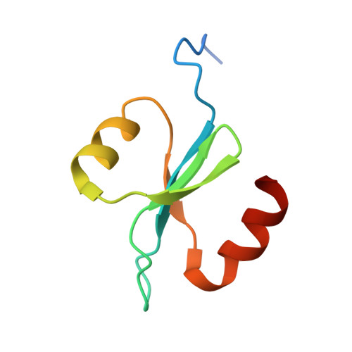
High avidity drives the interaction between the streptococcal C1 phage endolysin, PlyC, with the cell surface carbohydrates of Group A Streptococcus.
Broendum, S.S., Williams, D.E., Hayes, B.K., Kraus, F., Fodor, J., Clifton, B.E., Geert Volbeda, A., Codee, J.D.C., Riley, B.T., Drinkwater, N., Farrow, K.A., Tsyganov, K., Heselpoth, R.D., Nelson, D.C., Jackson, C.J., Buckle, A.M., McGowan, S.(2021) Mol Microbiol 116: 397-415
- PubMed: 33756056
- DOI: https://doi.org/10.1111/mmi.14719
- Primary Citation of Related Structures:
7KWT, 7KWW, 7KWY - PubMed Abstract:
Endolysin enzymes from bacteriophage cause bacterial lysis by degrading the peptidoglycan cell wall. The streptococcal C1 phage endolysin PlyC, is the most potent endolysin described to date and can rapidly lyse group A, C, and E streptococci. PlyC is known to bind the Group A streptococcal cell wall, but the specific molecular target or the binding site within PlyC remain uncharacterized. Here we report for the first time, that the polyrhamnose backbone of the Group A streptococcal cell wall is the binding target of PlyC. We have also characterized the putative rhamnose binding groove of PlyC and found four key residues that were critical to either the folding or the cell wall binding action of PlyC. Based on our results, we suggest that the interaction between PlyC and the cell wall may not be a high-affinity interaction as previously proposed, but rather a high avidity one, allowing for PlyC's remarkable lytic activity. Resistance to our current antibiotics is reaching crisis levels and there is an urgent need to develop the antibacterial agents with new modes of action. A detailed understanding of this potent endolysin may facilitate future developments of PlyC as a tool against the rise of antibiotic resistance.
Organizational Affiliation:
Biomedicine Discovery Institute, Department of Microbiology, Monash University, Melbourne, VIC, Australia.


