1OI6

Structure determination of the TMP-complex of EvaD


Experimental Data Snapshot

  • Method: X-RAY DIFFRACTION
  • Resolution: 1.40 Å
  • R-Value Free: 
    0.166 (Depositor), 0.180 (DCC) 
  • R-Value Work: 
    0.129 (Depositor), 0.140 (DCC) 
  • R-Value Observed: 
    0.130 (Depositor) 

Starting Model: experimental
View more details

wwPDB Validation   3D Report Full Report


Ligand Structure Quality Assessment 

Created with Raphaël 2.3.0Worse 01 BetterLigand structure goodness of fit to experimental dataBest fitted TMPClick on this verticalbar to view details

This is version 1.4 of the entry. See complete history


Literature

The Position of a Key Tyrosine in Dtdp-4-Keto-6-Deoxy-D-Glucose-5-Epimerase (Evad) Alters the Substrate Profile for This Rmlc-Like Enzyme

Merkel, A.B.Major, L.L.Errey, J.C.Burkart, M.D.Field, R.A.Walsh, C.T.Naismith, J.H.

(2004) J Biological Chem 279: 32684

  • DOI: https://doi.org/10.1074/jbc.M404091200
  • Primary Citation of Related Structures:  
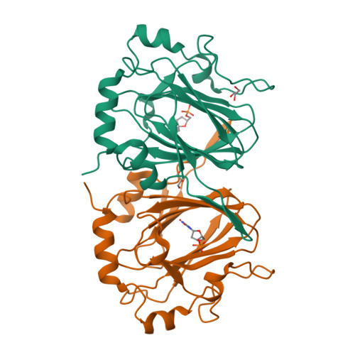
    1OI6

  • PubMed Abstract: 

    Vancomycin, the last line of defense antibiotic, depends upon the attachment of the carbohydrate vancosamine to an aglycone skeleton for antibacterial activity. Vancomycin is a naturally occurring secondary metabolite that can be produced by bacterial fermentation. To combat emerging resistance, it has been proposed to genetically engineer bacteria to produce analogues of vancomycin. This requires a detailed understanding of the biochemical steps in the synthesis of vancomycin. Here we report the 1.4 A structure and biochemical characterization of EvaD, an RmlC-like protein that is required for the C-5' epimerization during synthesis of dTDP-epivancosamine. EvaD, although clearly belonging to the RmlC class of enzymes, displays very low activity in the archetypal RmlC reaction (double epimerization of dTDP-6-deoxy-4-keto-D-glucose at C-3' and C-5'). The high resolution structure of EvaD compared with the structures of authentic RmlC enzymes indicates that a subtle change in the enzyme active site repositions a key catalytic Tyr residue. A mutant designed to re-establish the normal position of the Tyr increases the RmlC-like activity of EvaD.


  • Organizational Affiliation

    Centre for Biomolecular Sciences, The University, St. Andrews, Scotland, KY16 9ST, United Kingdom.


Macromolecules
Find similar proteins by:  (by identity cutoff)  |  3D Structure
Entity ID: 1
MoleculeChains Sequence LengthOrganismDetailsImage
PCZA361.16
A, B
205Amycolatopsis orientalisMutation(s): 0 
UniProt
Find proteins for O52806 (Amycolatopsis orientalis)
Explore O52806 
Go to UniProtKB:  O52806
Entity Groups  
Sequence Clusters30% Identity50% Identity70% Identity90% Identity95% Identity100% Identity
UniProt GroupO52806
Sequence Annotations
Expand
  • Reference Sequence
Experimental Data & Validation

Experimental Data

  • Method: X-RAY DIFFRACTION
  • Resolution: 1.40 Å
  • R-Value Free:  0.166 (Depositor), 0.180 (DCC) 
  • R-Value Work:  0.129 (Depositor), 0.140 (DCC) 
  • R-Value Observed: 0.130 (Depositor) 
Space Group: P 21 21 2
Unit Cell:
Length ( Å )Angle ( ˚ )
a = 98.5α = 90
b = 72.571β = 90
c = 57.239γ = 90
Software Package:
Software NamePurpose
REFMACrefinement
MOSFLMdata reduction
SCALEPACKdata scaling
AMoREphasing

Structure Validation

View Full Validation Report


Ligand Structure Quality Assessment 

Created with Raphaël 2.3.0Worse 01 BetterLigand structure goodness of fit to experimental dataBest fitted TMPClick on this verticalbar to view details

Entry History 

Deposition Data

Revision History  (Full details and data files)

  • Version 1.0: 2004-06-03
    Type: Initial release
  • Version 1.1: 2011-05-08
    Changes: Version format compliance
  • Version 1.2: 2011-07-13
    Changes: Version format compliance
  • Version 1.3: 2019-05-08
    Changes: Data collection, Experimental preparation, Other
  • Version 1.4: 2023-12-13
    Changes: Data collection, Database references, Other, Refinement description