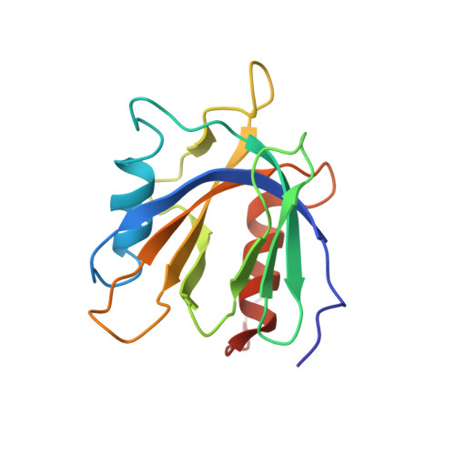
The PTB domain of tensin: NMR solution structure and phosphoinositides binding studies.
Leone, M., Yu, E.C., Liddington, R.C., Pasquale, E.B., Pellecchia, M.(2007) Biopolymers 89: 86-92
- PubMed: 17922498
- DOI: https://doi.org/10.1002/bip.20862
- Primary Citation of Related Structures:
2GJY - PubMed Abstract:
Tensin is a protein confined at those discrete and specialized regions of the plasma membrane, known as focal adhesions. It contains, at the C-terminus, a phosphotyrosine binding (PTB) domain that can interact with the cytoplasmic tail of beta-integrins and is necessary for localization of the protein to cell-matrix adhesions. Here, we present the NMR solution structure of the PTB domain of tensin1. Moreover, through NMR binding studies, we demonstrate that the PTB domain of tensin1 is able to interact with phosphatidylinositol 4, 5-diphosphate (PtIns(4,5)P2) and phosphatidylinositol 4-phosphate (PtIns(4)P), presenting higher affinity for the diphosphorylated inositide. Chemical shift mapping studies reveal a putative PtIns(4,5)P2 binding region that is distinct from the predicted integrin beta-tail recognition site. Heteronuclear NOE experiments, recorded in absence and presence of PtIns(4,5)P2, indicate that the interaction with lipids decreases the flexibility of loop regions, predicted to be important for integrin binding, and thus, proposes a possible correlation between the two distinct binding events. Therefore, our studies suggest that capture of lipids by the PTB domain of tensin1 may play a role for the protein function at focal adhesions.
Organizational Affiliation:
Cancer Center and Infectious and Inflammatory Disease Center, Burnham Institute for Medical Research, La Jolla, CA, USA.