3HYF

Crystal structure of HIV-1 RNase H p15 with engineered E. coli loop and active site inhibitor


Experimental Data Snapshot

  • Method: X-RAY DIFFRACTION
  • Resolution: 1.70 Å
  • R-Value Free: 
    0.232 (Depositor), 0.250 (DCC) 
  • R-Value Work: 
    0.196 (Depositor), 0.210 (DCC) 
  • R-Value Observed: 
    0.198 (Depositor) 

Starting Model: experimental
View more details

wwPDB Validation   3D Report Full Report


Ligand Structure Quality Assessment 

Created with Raphaël 2.3.0Worse 01 BetterLigand structure goodness of fit to experimental dataBest fitted ON1Click on this verticalbar to view details

This is version 1.5 of the entry. See complete history


Literature

RNase H active site inhibitors of human immunodeficiency virus type 1 reverse transcriptase: design, biochemical activity, and structural information.

Kirschberg, T.A.Balakrishnan, M.Squires, N.H.Barnes, T.Brendza, K.M.Chen, X.Eisenberg, E.J.Jin, W.Kutty, N.Leavitt, S.Liclican, A.Liu, Q.Liu, X.Mak, J.Perry, J.K.Wang, M.Watkins, W.J.Lansdon, E.B.

(2009) J Med Chem 52: 5781-5784

  • DOI: https://doi.org/10.1021/jm900597q
  • Primary Citation of Related Structures:  
    3HYF

  • PubMed Abstract: 

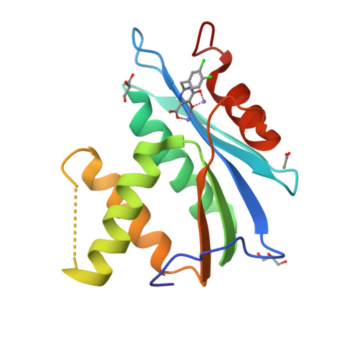
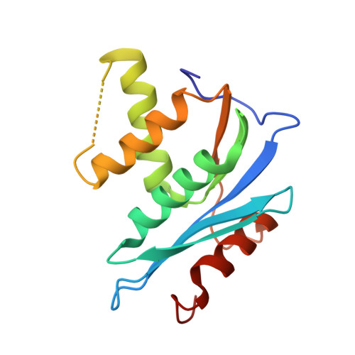
    Pyrimidinol carboxylic acids were designed as inhibitors of HIV-1 RNase H function. These molecules can coordinate to two divalent metal ions in the RNase H active site. Inhibition of enzymatic activity was measured in a biochemical assay, but no antiviral effect was observed. Binding was demonstrated via a solid state structure of the isolated p15-Ec domain of HIV-1 RT showing inhibitor and two Mn(II) ions bound to the RNase H active site.


  • Organizational Affiliation

    Department of Medicinal Chemistry, Gilead Sciences, Foster City, California 94404, USA. tkirschberg@gilead.com


Macromolecules
Find similar proteins by:  (by identity cutoff)  |  3D Structure
Entity ID: 1
MoleculeChains Sequence LengthOrganismDetailsImage
Reverse transcriptase/RNaseH150Human immunodeficiency virus 1Escherichia coli K-12Mutation(s): 0 
Gene Names: polReverse Transcriptase (amino acids 427-560)
EC: 3.1.26.4
UniProt
Find proteins for P0A7Y4 (Escherichia coli (strain K12))
Explore P0A7Y4 
Go to UniProtKB:  P0A7Y4
Find proteins for Q72547 (Human immunodeficiency virus type 1)
Explore Q72547 
Go to UniProtKB:  Q72547
Entity Groups  
Sequence Clusters30% Identity50% Identity70% Identity90% Identity95% Identity100% Identity
UniProt GroupsQ72547P0A7Y4
Sequence Annotations
Expand
  • Reference Sequence
Small Molecules
Ligands 4 Unique
IDChains Name / Formula / InChI Key2D Diagram3D Interactions
ON1
Query on ON1

Download Ideal Coordinates CCD File 
G [auth A]2-(3,4-dichlorobenzyl)-5,6-dihydroxypyrimidine-4-carboxylic acid
C12 H8 Cl2 N2 O4
OZDSTVKQUHMIRA-UHFFFAOYSA-N
GOL
Query on GOL

Download Ideal Coordinates CCD File 
E [auth A],
F [auth A]
GLYCEROL
C3 H8 O3
PEDCQBHIVMGVHV-UHFFFAOYSA-N
ACT
Query on ACT

Download Ideal Coordinates CCD File 
D [auth A]ACETATE ION
C2 H3 O2
QTBSBXVTEAMEQO-UHFFFAOYSA-M
MN
Query on MN

Download Ideal Coordinates CCD File 
B [auth A],
C [auth A]
MANGANESE (II) ION
Mn
WAEMQWOKJMHJLA-UHFFFAOYSA-N
Experimental Data & Validation

Experimental Data

  • Method: X-RAY DIFFRACTION
  • Resolution: 1.70 Å
  • R-Value Free:  0.232 (Depositor), 0.250 (DCC) 
  • R-Value Work:  0.196 (Depositor), 0.210 (DCC) 
  • R-Value Observed: 0.198 (Depositor) 
Space Group: I 21 21 21
Unit Cell:
Length ( Å )Angle ( ˚ )
a = 39.717α = 90
b = 90.326β = 90
c = 113.334γ = 90
Software Package:
Software NamePurpose
BOSdata collection
AMoREphasing
CNXrefinement
HKL-2000data reduction
HKL-2000data scaling

Structure Validation

View Full Validation Report


Ligand Structure Quality Assessment 

Created with Raphaël 2.3.0Worse 01 BetterLigand structure goodness of fit to experimental dataBest fitted ON1Click on this verticalbar to view details

Entry History 

Deposition Data

Revision History  (Full details and data files)

  • Version 1.0: 2009-10-20
    Type: Initial release
  • Version 1.1: 2011-07-13
    Changes: Non-polymer description, Version format compliance
  • Version 1.2: 2011-11-16
    Changes: Atomic model
  • Version 1.3: 2017-08-16
    Changes: Refinement description, Source and taxonomy
  • Version 1.4: 2023-09-06
    Changes: Data collection, Database references, Derived calculations, Refinement description
  • Version 1.5: 2024-03-13
    Changes: Source and taxonomy