


Crystal structure of abscisic acid-bound PYL10
Sun, D.M., Wu, M.H., Wang, H.P., Zang, J.Y., Tian, C.L.To be published.
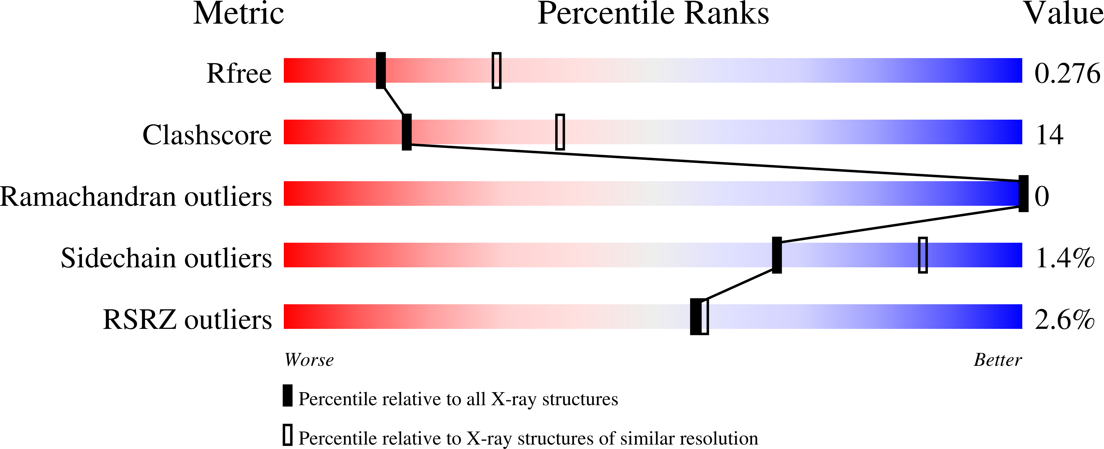
Experimental Data Snapshot
Starting Model: experimental
View more details
Entity ID: 1 | |||||
|---|---|---|---|---|---|
| Molecule | Chains | Sequence Length | Organism | Details | Image |
| Abscisic acid receptor PYL10 | 191 | Arabidopsis thaliana | Mutation(s): 0 Gene Names: PYL10 | 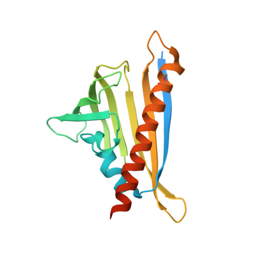 | |
UniProt | |||||
Find proteins for Q8H1R0 (Arabidopsis thaliana) Explore Q8H1R0 Go to UniProtKB: Q8H1R0 | |||||
Entity Groups | |||||
| Sequence Clusters | 30% Identity50% Identity70% Identity90% Identity95% Identity100% Identity | ||||
| UniProt Group | Q8H1R0 | ||||
Sequence AnnotationsExpand | |||||
| |||||
| Ligands 1 Unique | |||||
|---|---|---|---|---|---|
| ID | Chains | Name / Formula / InChI Key | 2D Diagram | 3D Interactions | |
| A8S Query on A8S | B [auth A] | (2Z,4E)-5-[(1S)-1-hydroxy-2,6,6-trimethyl-4-oxocyclohex-2-en-1-yl]-3-methylpenta-2,4-dienoic acid C15 H20 O4 JLIDBLDQVAYHNE-YKALOCIXSA-N |  | ||
| Length ( Å ) | Angle ( ˚ ) |
|---|---|
| a = 68.327 | α = 90 |
| b = 68.327 | β = 90 |
| c = 63.396 | γ = 120 |
| Software Name | Purpose |
|---|---|
| HKL-2000 | data collection |
| PHASES | phasing |
| REFMAC | refinement |
| HKL-2000 | data reduction |
| HKL-2000 | data scaling |
RCSB PDB is hosted by

RCSB PDB is a member of the

All questions and comments will receive a response in a timely manner.


