3UAS

Cytochrome P450 2B4 covalently bound to the mechanism-based inactivator 9-ethynylphenanthrene


Experimental Data Snapshot

  • Method: X-RAY DIFFRACTION
  • Resolution: 2.94 Å
  • R-Value Free: 
    0.296 (Depositor), 0.280 (DCC) 
  • R-Value Work: 
    0.243 (Depositor), 0.240 (DCC) 
  • R-Value Observed: 
    0.245 (Depositor) 

Starting Model: experimental
View more details

wwPDB Validation   3D Report Full Report


Ligand Structure Quality Assessment 

Created with Raphaël 2.3.0Worse 01 BetterLigand structure goodness of fit to experimental dataBest fitted HEMClick on this verticalbar to view detailsBest fitted CM5Click on this verticalbar to view detailsBest fitted 0BVClick on this verticalbar to view details

This is version 1.4 of the entry. See complete history


Literature

Potent Mechanism-Based Inactivation of Cytochrome P450 2B4 by 9-Ethynylphenanthrene: Implications for Allosteric Modulation of Cytochrome P450 Catalysis.

Zhang, H.Gay, S.C.Shah, M.Foroozesh, M.Liu, J.Osawa, Y.Zhang, Q.Stout, C.D.Halpert, J.R.Hollenberg, P.F.

(2013) Biochemistry 52: 355-364

  • DOI: https://doi.org/10.1021/bi301567z
  • Primary Citation of Related Structures:  
    3UAS

  • PubMed Abstract: 

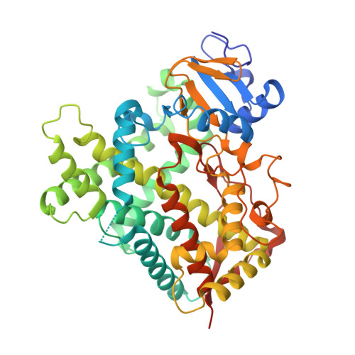
    The mechanism-based inactivation of cytochrome P450 2B4 (CYP2B4) by 9-ethynylphenanthrene (9EP) has been investigated. The partition ratio and k(inact) are 0.2 and 0.25 min(-1), respectively. Intriguingly, the inactivation exhibits sigmoidal kinetics with a Hill coefficient of 2.5 and an S(50) of 4.5 μM indicative of homotropic cooperativity. Enzyme inactivation led to an increase in mass of the apo-CYP2B4 by 218 Da as determined by electrospray ionization liquid chromatography and mass spectrometry, consistent with covalent protein modification. The modified CYP2B4 was purified to homogeneity and its structure determined by X-ray crystallography. The structure showed that 9EP is covalently attached to Oγ of Thr 302 via an ester bond, which is consistent with the increased mass of the protein. The presence of the bulky phenanthrenyl ring resulted in inward rotations of Phe 297 and Phe 206, leading to a compact active site. Thus, binding of another molecule of 9EP in the active site is prohibited. However, results from the quenching of 9EP fluorescence by unmodified or 9EP-modified CYP2B4 revealed at least two binding sites with distinct affinities, with the low-affinity site being the catalytic site and the high-affinity site on the protein periphery. Computer-aided docking and molecular dynamics simulations with one or two ligands bound revealed that the high-affinity site is situated at the entrance of a substrate access channel surrounded by the F' helix, β1-β2 loop, and β4 loop and functions as an allosteric site to enhance the efficiency of activation of the acetylenic group of 9EP and subsequent covalent modification of Thr 302.


  • Organizational Affiliation

    Department of Pharmacology, The University of Michigan Medical School , Ann Arbor, Michigan 48109, USA.


Macromolecules
Find similar proteins by:  (by identity cutoff)  |  3D Structure
Entity ID: 1
MoleculeChains Sequence LengthOrganismDetailsImage
Cytochrome P450 2B4476Oryctolagus cuniculusMutation(s): 9 
Gene Names: CYP2B4
EC: 1.14.14.1
UniProt
Find proteins for P00178 (Oryctolagus cuniculus)
Explore P00178 
Go to UniProtKB:  P00178
Entity Groups  
Sequence Clusters30% Identity50% Identity70% Identity90% Identity95% Identity100% Identity
UniProt GroupP00178
Sequence Annotations
Expand
  • Reference Sequence
Small Molecules
Ligands 4 Unique
IDChains Name / Formula / InChI Key2D Diagram3D Interactions
HEM
Query on HEM

Download Ideal Coordinates CCD File 
B [auth A]PROTOPORPHYRIN IX CONTAINING FE
C34 H32 Fe N4 O4
KABFMIBPWCXCRK-RGGAHWMASA-L
CM5
Query on CM5

Download Ideal Coordinates CCD File 
E [auth A]5-CYCLOHEXYL-1-PENTYL-BETA-D-MALTOSIDE
C23 H42 O11
RVTGFZGNOSKUDA-ZNGNCRBCSA-N
0BV
Query on 0BV

Download Ideal Coordinates CCD File 
C [auth A]phenanthren-9-ylacetaldehyde
C16 H12 O
GLWZSGUBBSQBKG-UHFFFAOYSA-N
TAM
Query on TAM

Download Ideal Coordinates CCD File 
D [auth A],
F [auth A]
TRIS(HYDROXYETHYL)AMINOMETHANE
C7 H17 N O3
GKODZWOPPOTFGA-UHFFFAOYSA-N
Experimental Data & Validation

Experimental Data

  • Method: X-RAY DIFFRACTION
  • Resolution: 2.94 Å
  • R-Value Free:  0.296 (Depositor), 0.280 (DCC) 
  • R-Value Work:  0.243 (Depositor), 0.240 (DCC) 
  • R-Value Observed: 0.245 (Depositor) 
Space Group: P 31 2 1
Unit Cell:
Length ( Å )Angle ( ˚ )
a = 90.155α = 90
b = 90.155β = 90
c = 148.623γ = 120
Software Package:
Software NamePurpose
SAINTdata scaling
XPREPdata reduction
PHASERphasing
REFMACrefinement
PDB_EXTRACTdata extraction
PROTEUM PLUSdata collection
SAINTdata reduction

Structure Validation

View Full Validation Report


Ligand Structure Quality Assessment 

Created with Raphaël 2.3.0Worse 01 BetterLigand structure goodness of fit to experimental dataBest fitted HEMClick on this verticalbar to view detailsBest fitted CM5Click on this verticalbar to view detailsBest fitted 0BVClick on this verticalbar to view details

Entry History 

Deposition Data

Revision History  (Full details and data files)

  • Version 1.0: 2013-01-09
    Type: Initial release
  • Version 1.1: 2013-01-30
    Changes: Database references
  • Version 1.2: 2018-01-24
    Changes: Structure summary
  • Version 1.3: 2023-09-13
    Changes: Data collection, Database references, Derived calculations, Refinement description
  • Version 1.4: 2024-11-27
    Changes: Structure summary