3PCR

Structure of EspG-Arf6 complex


Experimental Data Snapshot

  • Method: X-RAY DIFFRACTION
  • Resolution: 2.50 Å
  • R-Value Free: 
    0.326 (Depositor), 0.321 (DCC) 
  • R-Value Work: 
    0.223 (Depositor), 0.218 (DCC) 
  • R-Value Observed: 
    0.228 (Depositor) 

wwPDB Validation   3D Report Full Report


Ligand Structure Quality Assessment 

Created with Raphaël 2.3.0Worse 01 BetterLigand structure goodness of fit to experimental dataBest fitted GTPClick on this verticalbar to view details

This is version 1.2 of the entry. See complete history


Literature

The assembly of a GTPase-kinase signalling complex by a bacterial catalytic scaffold.

Selyunin, A.S.Sutton, S.E.Weigele, B.A.Reddick, L.E.Orchard, R.C.Bresson, S.M.Tomchick, D.R.Alto, N.M.

(2011) Nature 469: 107-111

  • DOI: https://doi.org/10.1038/nature09593
  • Primary Citation of Related Structures:  
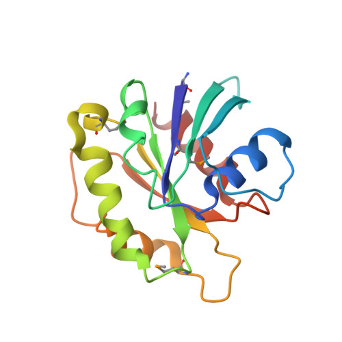
    3PCR, 3PCS

  • PubMed Abstract: 

    The fidelity and specificity of information flow within a cell is controlled by scaffolding proteins that assemble and link enzymes into signalling circuits. These circuits can be inhibited by bacterial effector proteins that post-translationally modify individual pathway components. However, there is emerging evidence that pathogens directly organize higher-order signalling networks through enzyme scaffolding, and the identity of the effectors and their mechanisms of action are poorly understood. Here we identify the enterohaemorrhagic Escherichia coli O157:H7 type III effector EspG as a regulator of endomembrane trafficking using a functional screen, and report ADP-ribosylation factor (ARF) GTPases and p21-activated kinases (PAKs) as its relevant host substrates. The 2.5 Å crystal structure of EspG in complex with ARF6 shows how EspG blocks GTPase-activating-protein-assisted GTP hydrolysis, revealing a potent mechanism of GTPase signalling inhibition at organelle membranes. In addition, the 2.8 Å crystal structure of EspG in complex with the autoinhibitory Iα3-helix of PAK2 defines a previously unknown catalytic site in EspG and provides an allosteric mechanism of kinase activation by a bacterial effector. Unexpectedly, ARF and PAKs are organized on adjacent surfaces of EspG, indicating its role as a 'catalytic scaffold' that effectively reprograms cellular events through the functional assembly of GTPase-kinase signalling complex.


  • Organizational Affiliation

    Department of Microbiology, University of Texas Southwestern Medical Center, 5323 Harry Hines Boulevard, Dallas, Texas 75390-8816, USA.


Macromolecules
Find similar proteins by:  (by identity cutoff)  |  3D Structure
Entity ID: 1
MoleculeChains Sequence LengthOrganismDetailsImage
EspG357Escherichia coli O157:H7Mutation(s): 0 
Gene Names: ECs4590espGZ5142
UniProt
Find proteins for Q7DB50 (Escherichia coli O157:H7)
Explore Q7DB50 
Go to UniProtKB:  Q7DB50
Entity Groups  
Sequence Clusters30% Identity50% Identity70% Identity90% Identity95% Identity100% Identity
UniProt GroupQ7DB50
Sequence Annotations
Expand
  • Reference Sequence
Find similar proteins by:  (by identity cutoff)  |  3D Structure
Entity ID: 2
MoleculeChains Sequence LengthOrganismDetailsImage
ADP-ribosylation factor 6162Homo sapiensMutation(s): 0 
Gene Names: ARF6
EC: 3.6.5.2
UniProt & NIH Common Fund Data Resources
Find proteins for P62330 (Homo sapiens)
Explore P62330 
Go to UniProtKB:  P62330
PHAROS:  P62330
GTEx:  ENSG00000165527 
Entity Groups  
Sequence Clusters30% Identity50% Identity70% Identity90% Identity95% Identity100% Identity
UniProt GroupP62330
Sequence Annotations
Expand
  • Reference Sequence
Small Molecules
Modified Residues  1 Unique
IDChains TypeFormula2D DiagramParent
MSE
Query on MSE
A
L-PEPTIDE LINKINGC5 H11 N O2 SeMET
Experimental Data & Validation

Experimental Data

  • Method: X-RAY DIFFRACTION
  • Resolution: 2.50 Å
  • R-Value Free:  0.326 (Depositor), 0.321 (DCC) 
  • R-Value Work:  0.223 (Depositor), 0.218 (DCC) 
  • R-Value Observed: 0.228 (Depositor) 
Space Group: P 43 21 2
Unit Cell:
Length ( Å )Angle ( ˚ )
a = 104.566α = 90
b = 104.566β = 90
c = 98.238γ = 90
Software Package:
Software NamePurpose
SCALEPACKdata scaling
PHENIXrefinement
PDB_EXTRACTdata extraction
HKL-3000data collection
HKL-3000data reduction
MLPHAREphasing

Structure Validation

View Full Validation Report


Ligand Structure Quality Assessment 

Created with Raphaël 2.3.0Worse 01 BetterLigand structure goodness of fit to experimental dataBest fitted GTPClick on this verticalbar to view details

Entry History 

Deposition Data

Revision History  (Full details and data files)

  • Version 1.0: 2011-01-05
    Type: Initial release
  • Version 1.1: 2011-07-13
    Changes: Source and taxonomy, Version format compliance
  • Version 1.2: 2024-10-30
    Changes: Data collection, Database references, Derived calculations, Structure summary