5TKF

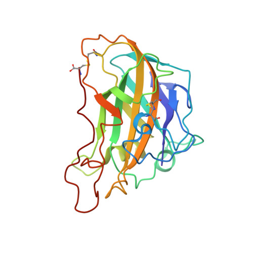
Neurospora crassa polysaccharide monooxygenase 2 high mannosylation


Experimental Data Snapshot

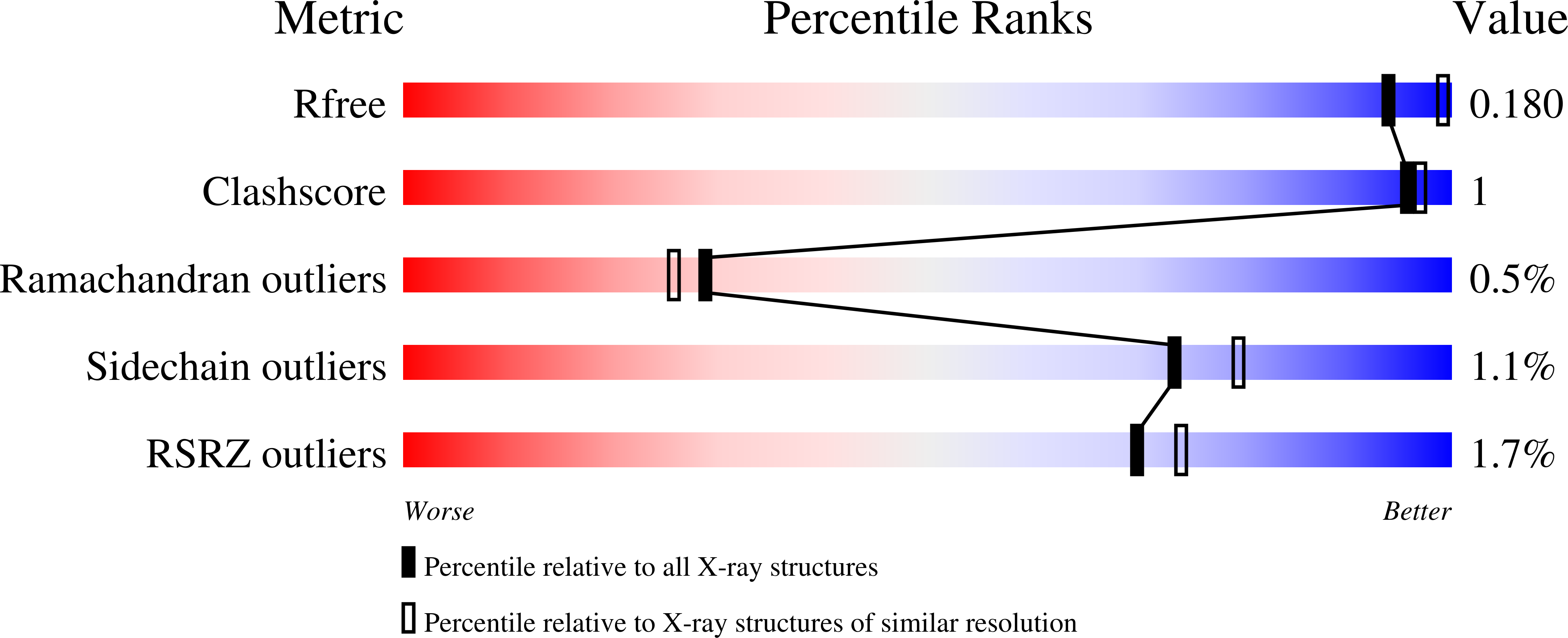
  • Method: X-RAY DIFFRACTION
  • Resolution: 2.10 Å
  • R-Value Free: 0.177 
  • R-Value Work: 0.142 
  • R-Value Observed: 0.143 

Starting Model: experimental
View more details

wwPDB Validation   3D Report Full Report


Ligand Structure Quality Assessment 


This is version 2.2 of the entry. See complete history


Literature

Crystallization of a fungal lytic polysaccharide monooxygenase expressed from glycoengineered Pichia pastoris for X-ray and neutron diffraction.

O'Dell, W.B.Swartz, P.D.Weiss, K.L.Meilleur, F.

(2017) Acta Crystallogr F Struct Biol Commun 73: 70-78

  • DOI: https://doi.org/10.1107/S2053230X16020318
  • Primary Citation of Related Structures:  
    5TKF

  • PubMed Abstract: 

    Lytic polysaccharide monooxygenases (LPMOs) are carbohydrate-disrupting enzymes secreted by bacteria and fungi that break glycosidic bonds via an oxidative mechanism. Fungal LPMOs typically act on cellulose and can enhance the efficiency of cellulose-hydrolyzing enzymes that release soluble sugars for bioethanol production or other industrial uses. The enzyme PMO-2 from Neurospora crassa (NcPMO-2) was heterologously expressed in Pichia pastoris to facilitate crystallographic studies of the fungal LPMO mechanism. Diffraction resolution and crystal morphology were improved by expressing NcPMO-2 from a glycoengineered strain of P. pastoris and by the use of crystal seeding methods, respectively. These improvements resulted in high-resolution (1.20 Å) X-ray diffraction data collection at 100 K and the production of a large NcPMO-2 crystal suitable for room-temperature neutron diffraction data collection to 2.12 Å resolution.


  • Organizational Affiliation

    Department of Molecular and Structural Biochemistry, North Carolina State University, Campus Box 7622, Raleigh, NC 27695, USA.


Macromolecules
Find similar proteins by:  (by identity cutoff)  |  3D Structure
Entity ID: 1
MoleculeChains Sequence LengthOrganismDetailsImage
Lytic polysaccharide monooxygenase
A, B, C, D
223Neurospora crassaMutation(s): 0 
Gene Names: G15G9.090GE21DRAFT_7469
EC: 1.14.99.56
UniProt
Find proteins for Q1K8B6 (Neurospora crassa (strain ATCC 24698 / 74-OR23-1A / CBS 708.71 / DSM 1257 / FGSC 987))
Explore Q1K8B6 
Go to UniProtKB:  Q1K8B6
Entity Groups  
Sequence Clusters30% Identity50% Identity70% Identity90% Identity95% Identity100% Identity
UniProt GroupQ1K8B6
Glycosylation
Glycosylation Sites: 1
Sequence Annotations
Expand
  • Reference Sequence
Oligosaccharides

Help

Entity ID: 2
MoleculeChains Length2D Diagram Glycosylation3D Interactions
alpha-D-mannopyranose-(1-2)-alpha-D-mannopyranose-(1-3)-[alpha-D-mannopyranose-(1-3)-alpha-D-mannopyranose-(1-6)]alpha-D-mannopyranose-(1-4)-2-acetamido-2-deoxy-beta-D-glucopyranose-(1-4)-2-acetamido-2-deoxy-beta-D-glucopyranose
E
7N-Glycosylation
Glycosylation Resources
GlyTouCan:  G84676UY
GlyCosmos:  G84676UY
GlyGen:  G84676UY
Entity ID: 3
MoleculeChains Length2D Diagram Glycosylation3D Interactions
2-acetamido-2-deoxy-beta-D-glucopyranose-(1-4)-2-acetamido-2-deoxy-beta-D-glucopyranose
F
2N-Glycosylation
Glycosylation Resources
GlyTouCan:  G42666HT
GlyCosmos:  G42666HT
GlyGen:  G42666HT
Entity ID: 4
MoleculeChains Length2D Diagram Glycosylation3D Interactions
alpha-D-mannopyranose-(1-3)-alpha-D-mannopyranose-(1-4)-2-acetamido-2-deoxy-beta-D-glucopyranose-(1-4)-2-acetamido-2-deoxy-beta-D-glucopyranose
G
4N-Glycosylation
Glycosylation Resources
GlyTouCan:  G48068RF
GlyCosmos:  G48068RF
GlyGen:  G48068RF
Small Molecules
Ligands 3 Unique
IDChains Name / Formula / InChI Key2D Diagram3D Interactions
NAG
Query on NAG

Download Ideal Coordinates CCD File 
L [auth D]2-acetamido-2-deoxy-beta-D-glucopyranose
C8 H15 N O6
OVRNDRQMDRJTHS-FMDGEEDCSA-N
CU
Query on CU

Download Ideal Coordinates CCD File 
H [auth A],
J [auth B],
K [auth C],
M [auth D]
COPPER (II) ION
Cu
JPVYNHNXODAKFH-UHFFFAOYSA-N
OXY
Query on OXY

Download Ideal Coordinates CCD File 
I [auth B],
N [auth D]
OXYGEN MOLECULE
O2
MYMOFIZGZYHOMD-UHFFFAOYSA-N
Experimental Data & Validation

Experimental Data

  • Method: X-RAY DIFFRACTION
  • Resolution: 2.10 Å
  • R-Value Free: 0.177 
  • R-Value Work: 0.142 
  • R-Value Observed: 0.143 
  • Space Group: P 1
Unit Cell:
Length ( Å )Angle ( ˚ )
a = 43.3α = 97.57
b = 67.04β = 97.61
c = 84.15γ = 97.43
Software Package:
Software NamePurpose
PHENIXrefinement
MOSFLMdata reduction
Aimlessdata scaling
PHASERphasing
PDB_EXTRACTdata extraction

Structure Validation

View Full Validation Report


Ligand Structure Quality Assessment 


Entry History & Funding Information

Deposition Data


Funding OrganizationLocationGrant Number
National Science Foundation (NSF, United States)United States1069091

Revision History  (Full details and data files)

  • Version 1.0: 2017-05-17
    Type: Initial release
  • Version 1.1: 2017-09-20
    Changes: Author supporting evidence
  • Version 1.2: 2019-11-27
    Changes: Author supporting evidence
  • Version 2.0: 2020-07-29
    Type: Remediation
    Reason: Carbohydrate remediation
    Changes: Atomic model, Data collection, Derived calculations, Structure summary
  • Version 2.1: 2023-10-04
    Changes: Data collection, Database references, Derived calculations, Refinement description, Structure summary
  • Version 2.2: 2024-11-20
    Changes: Structure summary