7FH1

Structure of the human Meckelin


Experimental Data Snapshot

  • Method: ELECTRON MICROSCOPY
  • Resolution: 3.34 Å
  • Aggregation State: PARTICLE 
  • Reconstruction Method: SINGLE PARTICLE 

wwPDB Validation   3D Report Full Report


This is version 1.0 of the entry. See complete history


Literature

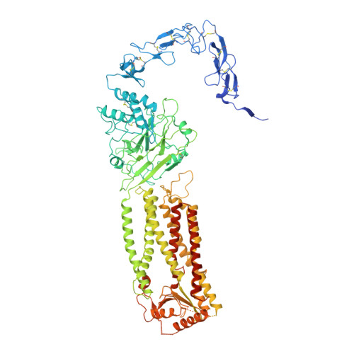
Structure of the human Meckel-Gruber protein Meckelin.

Liu, D.Qian, D.Shen, H.Gong, D.

(2021) Sci Adv 7: eabj9748-eabj9748

  • DOI: https://doi.org/10.1126/sciadv.abj9748
  • Primary Citation of Related Structures:  
    7FH1

  • PubMed Abstract: 

    Mutations in the Meckelin gene account for most cases of the Meckel-Gruber syndrome, the most severe ciliopathy with a 100% mortality rate. Here, we report a 3.3-Å cryo–electron microscopy structure of human Meckelin (also known as TMEM67 and MKS3). The structure reveals a unique protein fold consisting of an unusual cysteine-rich domain that folds as an arch bridge stabilized by 11 pairs of disulfide bonds, a previously uncharacterized domain named β sheet–rich domain, a previously unidentified seven-transmembrane fold wherein TM4 to TM6 are broken near the cytoplasmic surface of the membrane, and a coiled-coil domain placed below the transmembrane domain. Meckelin forms a stable homodimer with an extensive dimer interface. Our structure establishes a framework for dissecting the function and disease mechanisms of Meckelin.


  • Organizational Affiliation

    Key Laboratory of Structural Biology of Zhejiang Province, School of Life Sciences, Westlake University, Hangzhou, Zhejiang 310024, China.


Macromolecules
Find similar proteins by:  (by identity cutoff)  |  3D Structure
Entity ID: 1
MoleculeChains Sequence LengthOrganismDetailsImage
Meckelin
A, B
1,021Homo sapiensMutation(s): 0 
Gene Names: TMEM67MKS3
Membrane Entity: Yes 
UniProt & NIH Common Fund Data Resources
Find proteins for Q5HYA8 (Homo sapiens)
Explore Q5HYA8 
Go to UniProtKB:  Q5HYA8
PHAROS:  Q5HYA8
GTEx:  ENSG00000164953 
Entity Groups  
Sequence Clusters30% Identity50% Identity70% Identity90% Identity95% Identity100% Identity
UniProt GroupQ5HYA8
Sequence Annotations
Expand
  • Reference Sequence
Oligosaccharides

Help

Entity ID: 2
MoleculeChains Length2D Diagram Glycosylation3D Interactions
2-acetamido-2-deoxy-beta-D-glucopyranose-(1-4)-2-acetamido-2-deoxy-beta-D-glucopyranose
C, D, E, G, H
C, D, E, G, H, I
2N/A
Glycosylation Resources
GlyTouCan:  G42666HT
GlyCosmos:  G42666HT
GlyGen:  G42666HT
Entity ID: 3
MoleculeChains Length2D Diagram Glycosylation3D Interactions
beta-D-mannopyranose-(1-3)-[beta-D-mannopyranose-(1-6)]beta-D-mannopyranose-(1-4)-2-acetamido-2-deoxy-beta-D-glucopyranose-(1-4)-2-acetamido-2-deoxy-beta-D-glucopyranose
F, J
5N/A
Glycosylation Resources
GlyTouCan:  G89225LT
GlyCosmos:  G89225LT
GlyGen:  G89225LT
Experimental Data & Validation

Experimental Data

  • Method: ELECTRON MICROSCOPY
  • Resolution: 3.34 Å
  • Aggregation State: PARTICLE 
  • Reconstruction Method: SINGLE PARTICLE 

Structure Validation

View Full Validation Report


Entry History & Funding Information

Deposition Data

  • Released Date: 2021-12-01 
  • Deposition Author(s): Gong, D.S.

Funding OrganizationLocationGrant Number
Not funded--

Revision History  (Full details and data files)

  • Version 1.0: 2021-12-01
    Type: Initial release