8C3U

Crystal Structure of human IL-1beta in complex with a low molecular weight antagonist


Experimental Data Snapshot

  • Method: X-RAY DIFFRACTION
  • Resolution: 1.95 Å
  • R-Value Free: 
    0.241 (Depositor), 0.240 (DCC) 
  • R-Value Work: 
    0.208 (Depositor), 0.200 (DCC) 
  • R-Value Observed: 
    0.209 (Depositor) 

Starting Model: experimental
View more details

wwPDB Validation   3D Report Full Report


Ligand Structure Quality Assessment 

Created with Raphaël 2.3.0Worse 01 BetterLigand structure goodness of fit to experimental dataBest fitted T9CClick on this verticalbar to view details

This is version 1.1 of the entry. See complete history


Literature

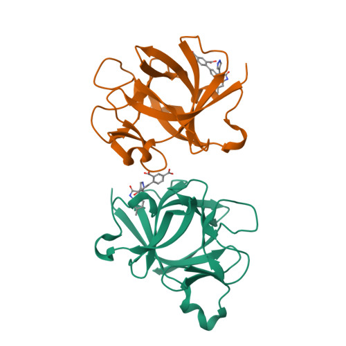
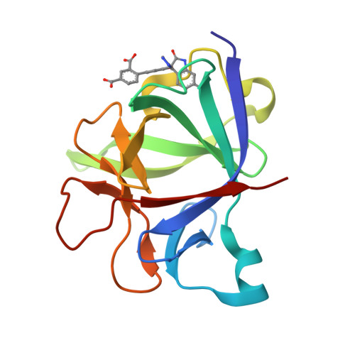
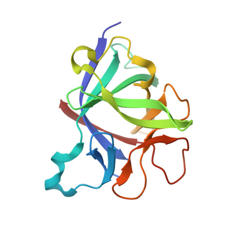
Discovery of a selective and biologically active low-molecular weight antagonist of human interleukin-1 beta.

Hommel, U.Hurth, K.Rondeau, J.M.Vulpetti, A.Ostermeier, D.Boettcher, A.Brady, J.P.Hediger, M.Lehmann, S.Koch, E.Blechschmidt, A.Yamamoto, R.Tundo Dottorello, V.Haenni-Holzinger, S.Kaiser, C.Lehr, P.Lingel, A.Mureddu, L.Schleberger, C.Blank, J.Ramage, P.Freuler, F.Eder, J.Bornancin, F.

(2023) Nat Commun 14: 5497-5497

  • DOI: https://doi.org/10.1038/s41467-023-41190-0
  • Primary Citation of Related Structures:  
    8C3U

  • PubMed Abstract: 

    Human interleukin-1β (hIL-1β) is a pro-inflammatory cytokine involved in many diseases. While hIL-1β directed antibodies have shown clinical benefit, an orally available low-molecular weight antagonist is still elusive, limiting the applications of hIL-1β-directed therapies. Here we describe the discovery of a low-molecular weight hIL-1β antagonist that blocks the interaction with the IL-1R1 receptor. Starting from a low affinity fragment-based screening hit 1, structure-based optimization resulted in a compound (S)-2 that binds and antagonizes hIL-1β with single-digit micromolar activity in biophysical, biochemical, and cellular assays. X-ray analysis reveals an allosteric mode of action that involves a hitherto unknown binding site in hIL-1β encompassing two loops involved in hIL-1R1/hIL-1β interactions. We show that residues of this binding site are part of a conformationally excited state of the mature cytokine. The compound antagonizes hIL-1β function in cells, including primary human fibroblasts, demonstrating the relevance of this discovery for future development of hIL-1β directed therapeutics.


  • Organizational Affiliation

    Novartis Institutes for BioMedical Research, Novartis Campus, CH-4002, Basel, Switzerland. uli.hommel@icloud.com.


Macromolecules
Find similar proteins by:  (by identity cutoff)  |  3D Structure
Entity ID: 1
MoleculeChains Sequence LengthOrganismDetailsImage
Interleukin-1 beta
A, B
153Homo sapiensMutation(s): 0 
Gene Names: IL1BIL1F2
UniProt & NIH Common Fund Data Resources
Find proteins for P01584 (Homo sapiens)
Explore P01584 
Go to UniProtKB:  P01584
PHAROS:  P01584
GTEx:  ENSG00000125538 
Entity Groups  
Sequence Clusters30% Identity50% Identity70% Identity90% Identity95% Identity100% Identity
UniProt GroupP01584
Sequence Annotations
Expand
  • Reference Sequence
Small Molecules
Ligands 1 Unique
IDChains Name / Formula / InChI Key2D Diagram3D Interactions
T9C (Subject of Investigation/LOI)
Query on T9C

Download Ideal Coordinates CCD File 
C [auth A],
D [auth B]
(S)-4'-hydroxy-3'-(6-methyl-2-oxo-3-(1H-pyrazol-4-yl)indolin-3-yl)-[1,1'-biphenyl]-2,4-dicarboxylic acid
C26 H19 N3 O6
QNQTUANCNUSMBX-AREMUKBSSA-N
Experimental Data & Validation

Experimental Data

  • Method: X-RAY DIFFRACTION
  • Resolution: 1.95 Å
  • R-Value Free:  0.241 (Depositor), 0.240 (DCC) 
  • R-Value Work:  0.208 (Depositor), 0.200 (DCC) 
  • R-Value Observed: 0.209 (Depositor) 
Space Group: P 1 21 1
Unit Cell:
Length ( Å )Angle ( ˚ )
a = 56.168α = 90
b = 60.085β = 114.76
c = 55.877γ = 90
Software Package:
Software NamePurpose
BUSTERrefinement
Aimlessdata scaling
XDSdata reduction
PHASERphasing

Structure Validation

View Full Validation Report


Ligand Structure Quality Assessment 

Created with Raphaël 2.3.0Worse 01 BetterLigand structure goodness of fit to experimental dataBest fitted T9CClick on this verticalbar to view details

Entry History & Funding Information

Deposition Data


Funding OrganizationLocationGrant Number
Not funded--

Revision History  (Full details and data files)

  • Version 1.0: 2023-09-13
    Type: Initial release
  • Version 1.1: 2024-03-27
    Changes: Database references