9FT9

Structure of the bipartite JNK1-JIP1 complex


Experimental Data Snapshot

  • Method: X-RAY DIFFRACTION
  • Resolution: 2.35 Å
  • R-Value Free: 
    0.270 (Depositor), 0.269 (DCC) 
  • R-Value Work: 
    0.231 (Depositor), 0.232 (DCC) 
  • R-Value Observed: 
    0.234 (Depositor) 

Starting Models: experimental
View more details

wwPDB Validation   3D Report Full Report


Ligand Structure Quality Assessment 

Created with Raphaël 2.3.0Worse 01 BetterLigand structure goodness of fit to experimental dataBest fitted ANPClick on this verticalbar to view details

This is version 1.2 of the entry. See complete history


Literature

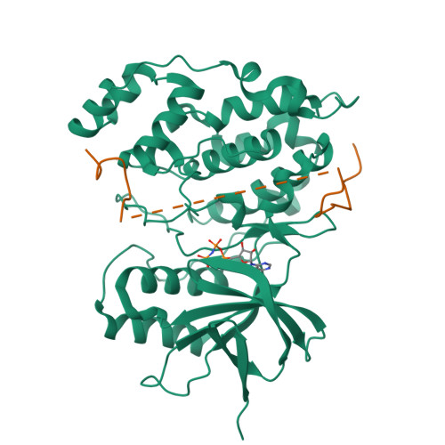
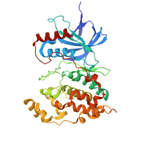
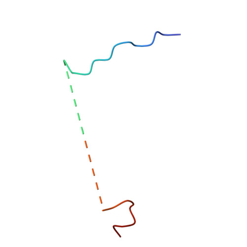
Bipartite binding of the intrinsically disordered scaffold protein JIP1 to the kinase JNK1.

Orand, T.Delaforge, E.Lee, A.Kragelj, J.Tengo, M.Tengo, L.Blackledge, M.Boeri Erba, E.Davis, R.J.Palencia, A.Jensen, M.R.

(2025) Proc Natl Acad Sci U S A 122: e2419915122-e2419915122

  • DOI: https://doi.org/10.1073/pnas.2419915122
  • Primary Citation of Related Structures:  
    9FT9

  • PubMed Abstract: 

    Scaffold proteins are key players in many signaling pathways where they ensure spatial and temporal control of molecular interactions by simultaneous tethering of multiple signaling components. The protein JIP1 acts as a scaffold within the c-Jun N-terminal kinase (JNK) signaling pathway by assembling three kinases, MLK3, MKK7, and JNK, into a macromolecular complex that enables their specific activation. The recruitment of these kinases depends on the 450-amino acid intrinsically disordered tail of JIP1, however, the structural details of this tail and the molecular mechanisms by which it binds kinases have remained elusive. Here, we provide an atomic resolution structural description of the JIP1 tail, and we study its interaction with the kinase JNK1. Using NMR spectroscopy, we show that JNK1 not only engages with the well-known docking site motif (D-motif) of JIP1, but also interacts with a noncanonical F-motif. We determine the crystal structure of the JIP1-JNK1 complex at 2.35 Å resolution revealing a bipartite binding mode of JIP1. Our work provides insights into the sequence determinants of F-motifs suggesting that these motifs may be more prevalent in JNK substrates than previously recognized. More broadly, our study highlights the power of NMR spectroscopy in uncovering kinase interaction motifs within disordered scaffold proteins, and it paves the way for atomic-resolution interaction studies of JIP1 with its multitude of interaction partners.


  • Organizational Affiliation

    Université Grenoble Alpes, Commissariat à l'Énergie Atomique et aux Énergies Alternatives, CNRS, Institut de Biologie Structurale, Grenoble 38044, France.


Macromolecules
Find similar proteins by:  (by identity cutoff)  |  3D Structure
Entity ID: 1
MoleculeChains Sequence LengthOrganismDetailsImage
Isoform 1 of Mitogen-activated protein kinase 8360Homo sapiensMutation(s): 0 
Gene Names: MAPK8JNK1PRKM8SAPK1SAPK1C
EC: 2.7.11.24
UniProt & NIH Common Fund Data Resources
Find proteins for P45983 (Homo sapiens)
Explore P45983 
Go to UniProtKB:  P45983
PHAROS:  P45983
GTEx:  ENSG00000107643 
Entity Groups  
Sequence Clusters30% Identity50% Identity70% Identity90% Identity95% Identity100% Identity
UniProt GroupP45983
Sequence Annotations
Expand
  • Reference Sequence
Find similar proteins by:  (by identity cutoff)  |  3D Structure
Entity ID: 2
MoleculeChains Sequence LengthOrganismDetailsImage
C-Jun-amino-terminal kinase-interacting protein 138Homo sapiensMutation(s): 0 
Gene Names: MAPK8IP1IB1JIP1PRKM8IP
Entity Groups  
Sequence Clusters30% Identity50% Identity70% Identity90% Identity95% Identity100% Identity
Sequence Annotations
Expand
  • Reference Sequence
Small Molecules
Ligands 1 Unique
IDChains Name / Formula / InChI Key2D Diagram3D Interactions
ANP (Subject of Investigation/LOI)
Query on ANP

Download Ideal Coordinates CCD File 
C [auth A]PHOSPHOAMINOPHOSPHONIC ACID-ADENYLATE ESTER
C10 H17 N6 O12 P3
PVKSNHVPLWYQGJ-KQYNXXCUSA-N
Experimental Data & Validation

Experimental Data

  • Method: X-RAY DIFFRACTION
  • Resolution: 2.35 Å
  • R-Value Free:  0.270 (Depositor), 0.269 (DCC) 
  • R-Value Work:  0.231 (Depositor), 0.232 (DCC) 
  • R-Value Observed: 0.234 (Depositor) 
Space Group: P 31 2 1
Unit Cell:
Length ( Å )Angle ( ˚ )
a = 137.7α = 90
b = 137.7β = 90
c = 63.95γ = 120
Software Package:
Software NamePurpose
PHENIXrefinement
XDSdata reduction
XSCALEdata scaling
PHASERphasing

Structure Validation

View Full Validation Report


Ligand Structure Quality Assessment 

Created with Raphaël 2.3.0Worse 01 BetterLigand structure goodness of fit to experimental dataBest fitted ANPClick on this verticalbar to view details

Entry History & Funding Information

Deposition Data


Funding OrganizationLocationGrant Number
Agence Nationale de la Recherche (ANR)FranceScaffoldDisorder
La ligue contre le cancerFranceThibault Orand thesis funding

Revision History  (Full details and data files)

  • Version 1.0: 2025-02-05
    Type: Initial release
  • Version 1.1: 2025-02-12
    Changes: Database references
  • Version 1.2: 2025-03-12
    Changes: Database references