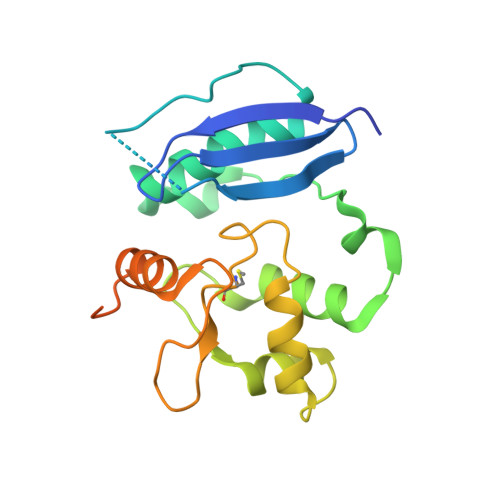
1EH7

METHYLATED HUMAN O6-ALKYLGUANINE-DNA ALKYLTRANSFERASE


Experimental Data Snapshot

  • Method: X-RAY DIFFRACTION
  • Resolution: 2.00 Å
  • R-Value Free: 0.229 
  • R-Value Work: 0.199 

Starting Model: experimental
View more details

wwPDB Validation   3D Report Full Report


This is version 1.4 of the entry. See complete history


Literature

Active and alkylated human AGT structures: a novel zinc site, inhibitor and extrahelical base binding.

Daniels, D.S.Mol, C.D.Arvai, A.S.Kanugula, S.Pegg, A.E.Tainer, J.A.

(2000) EMBO J 19: 1719-1730

  • DOI: https://doi.org/10.1093/emboj/19.7.1719
  • Primary Citation of Related Structures:  
    1EH6, 1EH7, 1EH8

  • PubMed Abstract: 

    Human O(6)-alkylguanine-DNA alkyltransferase (AGT), which directly reverses endogenous alkylation at the O(6)-position of guanine, confers resistance to alkylation chemotherapies and is therefore an active anticancer drug target. Crystal structures of active human AGT and its biologically and therapeutically relevant methylated and benzylated product complexes reveal an unexpected zinc-stabilized helical bridge joining a two-domain alpha/beta structure. An asparagine hinge couples the active site motif to a helix-turn-helix (HTH) motif implicated in DNA binding. The reactive cysteine environment, its position within a groove adjacent to the alkyl-binding cavity and mutational analyses characterize DNA-damage recognition and inhibitor specificity, support a structure-based dealkylation mechanism and suggest a molecular basis for destabilization of the alkylated protein. These results support damaged nucleotide flipping facilitated by an arginine finger within the HTH motif to stabilize the extrahelical O(6)-alkylguanine without the protein conformational change originally proposed from the empty Ada structure. Cysteine alkylation sterically shifts the HTH recognition helix to evidently mechanistically couple release of repaired DNA to an opening of the protein fold to promote the biological turnover of the alkylated protein.


  • Organizational Affiliation

    The Skaggs Institute for Chemical Biology, Department of Molecular Biology, MB-4, The Scripps Research Institute, 10550 North Torrey Pines Road, La Jolla, CA 92037-1027, USA.


Macromolecules
Find similar proteins by:  (by identity cutoff)  |  3D Structure
Entity ID: 1
MoleculeChains Sequence LengthOrganismDetailsImage
O6-ALKYLGUANINE-DNA ALKYLTRANSFERASE207Homo sapiensMutation(s): 0 
EC: 2.1.1.63
UniProt & NIH Common Fund Data Resources
Find proteins for P16455 (Homo sapiens)
Explore P16455 
Go to UniProtKB:  P16455
PHAROS:  P16455
GTEx:  ENSG00000170430 
Entity Groups  
Sequence Clusters30% Identity50% Identity70% Identity90% Identity95% Identity100% Identity
UniProt GroupP16455
Sequence Annotations
Expand
  • Reference Sequence
Small Molecules
Ligands 1 Unique
IDChains Name / Formula / InChI Key2D Diagram3D Interactions
ZN
Query on ZN

Download Ideal Coordinates CCD File 
B [auth A]ZINC ION
Zn
PTFCDOFLOPIGGS-UHFFFAOYSA-N
Modified Residues  1 Unique
IDChains TypeFormula2D DiagramParent
SMC
Query on SMC
A
L-PEPTIDE LINKINGC4 H9 N O2 SCYS
Experimental Data & Validation

Experimental Data

  • Method: X-RAY DIFFRACTION
  • Resolution: 2.00 Å
  • R-Value Free: 0.229 
  • R-Value Work: 0.199 
  • Space Group: P 31 2 1
Unit Cell:
Length ( Å )Angle ( ˚ )
a = 71.401α = 90
b = 71.401β = 90
c = 76.325γ = 120
Software Package:
Software NamePurpose
CNSrefinement
DENZOdata reduction
SCALEPACKdata scaling
CNSphasing

Structure Validation

View Full Validation Report


Entry History 

Deposition Data

Revision History  (Full details and data files)

  • Version 1.0: 2000-04-12
    Type: Initial release
  • Version 1.1: 2007-10-16
    Changes: Version format compliance
  • Version 1.2: 2011-07-13
    Changes: Version format compliance
  • Version 1.3: 2023-08-09
    Changes: Data collection, Database references, Derived calculations, Refinement description
  • Version 1.4: 2024-10-16
    Changes: Structure summary