1QZY

Human Methionine Aminopeptidase in complex with bengamide inhibitor LAF153 and cobalt


Experimental Data Snapshot

  • Method: X-RAY DIFFRACTION
  • Resolution: 1.60 Å
  • R-Value Free: 0.211 
  • R-Value Work: 0.204 
  • R-Value Observed: 0.204 

wwPDB Validation   3D Report Full Report


Ligand Structure Quality Assessment 


This is version 1.2 of the entry. See complete history


Literature

Proteomics-based target identification: bengamides as a new class of methionine aminopeptidase inhibitors.

Towbin, H.Bair, K.W.DeCaprio, J.A.Eck, M.J.Kim, S.Kinder, F.R.Morollo, A.Mueller, D.R.Schindler, P.Song, H.K.van Oostrum, J.Versace, R.W.Voshol, H.Wood, J.Zabludoff, S.Phillips, P.E.

(2003) J Biol Chem 278: 52964-52971

  • DOI: https://doi.org/10.1074/jbc.M309039200
  • Primary Citation of Related Structures:  
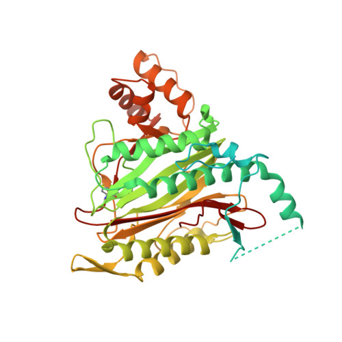
    1QZY

  • PubMed Abstract: 

    LAF389 is a synthetic analogue of bengamides, a class of marine natural products that produce inhibitory effects on tumor growth in vitro and in vivo. A proteomics-based approach has been used to identify signaling pathways affected by bengamides. LAF389 treatment of cells resulted in altered mobility of a subset of proteins on two-dimensional gel electrophoresis. Detailed analysis of one of the proteins, 14-3-3gamma, showed that bengamide treatment resulted in retention of the amino-terminal methionine, suggesting that bengamides directly or indirectly inhibited methionine aminopeptidases (MetAps). Both known MetAps are inhibited by LAF389. Short interfering RNA suppression of MetAp2 also altered amino-terminal processing of 14-3-3gamma. A high resolution structure of human MetAp2 co-crystallized with a bengamide shows that the compound binds in a manner that mimics peptide substrates. Additionally, the structure reveals that three key hydroxyl groups on the inhibitor coordinate the di-cobalt center in the enzyme active site.


  • Organizational Affiliation

    Novartis Pharma AG, CH-4036 Basle, Switzerland.


Macromolecules
Find similar proteins by:  (by identity cutoff)  |  3D Structure
Entity ID: 1
MoleculeChains Sequence LengthOrganismDetailsImage
Methionine aminopeptidase 2478Homo sapiensMutation(s): 0 
EC: 3.4.11.18
UniProt & NIH Common Fund Data Resources
Find proteins for P50579 (Homo sapiens)
Explore P50579 
Go to UniProtKB:  P50579
PHAROS:  P50579
GTEx:  ENSG00000111142 
Entity Groups  
Sequence Clusters30% Identity50% Identity70% Identity90% Identity95% Identity100% Identity
UniProt GroupP50579
Sequence Annotations
Expand
  • Reference Sequence
Small Molecules
Ligands 3 Unique
IDChains Name / Formula / InChI Key2D Diagram3D Interactions
TDE
Query on TDE

Download Ideal Coordinates CCD File 
D [auth A](E)-(2R,3R,4S,5R)-3,4,5-TRIHYDROXY-2-METHOXY-8,8-DIMETHYL-NON-6-ENOIC ACID ((3S,6R)-6-HYDROXY-2-OXO-AZEPAN-3-YL)-AMIDE
C18 H32 N2 O7
FVBBTOIQETYGOA-SDNORSGYSA-N
TBU
Query on TBU

Download Ideal Coordinates CCD File 
E [auth A]TERTIARY-BUTYL ALCOHOL
C4 H10 O
DKGAVHZHDRPRBM-UHFFFAOYSA-N
CO
Query on CO

Download Ideal Coordinates CCD File 
B [auth A],
C [auth A]
COBALT (II) ION
Co
XLJKHNWPARRRJB-UHFFFAOYSA-N
Binding Affinity Annotations 
IDSourceBinding Affinity
TDE Binding MOAD:  1QZY IC50: 800 (nM) from 1 assay(s)
Experimental Data & Validation

Experimental Data

  • Method: X-RAY DIFFRACTION
  • Resolution: 1.60 Å
  • R-Value Free: 0.211 
  • R-Value Work: 0.204 
  • R-Value Observed: 0.204 
  • Space Group: C 2 2 21
Unit Cell:
Length ( Å )Angle ( ˚ )
a = 90.61α = 90
b = 99.04β = 90
c = 101.383γ = 90
Software Package:
Software NamePurpose
CNSrefinement

Structure Validation

View Full Validation Report


Ligand Structure Quality Assessment 


Entry History 

Deposition Data

Revision History  (Full details and data files)

  • Version 1.0: 2003-11-25
    Type: Initial release
  • Version 1.1: 2008-04-29
    Changes: Version format compliance
  • Version 1.2: 2011-07-13
    Changes: Version format compliance