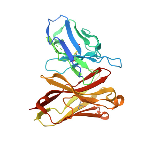
Substantial energetic improvement with minimal structural perturbation in a high affinity mutant antibody
Midelfort, K.S., Hernandez, H.H., Lippow, S.M., Tidor, B., Drennan, C.L., Wittrup, K.D.(2004) J Mol Biol 343: 685-701
- PubMed: 15465055
- DOI: https://doi.org/10.1016/j.jmb.2004.08.019
- Primary Citation of Related Structures:
1X9Q - PubMed Abstract:
Here, we compare an antibody with the highest known engineered affinity (K(d)=270 fM) to its high affinity wild-type (K(d)=700 pM) through thermodynamic, kinetic, structural, and theoretical analyses. The 4M5.3 anti-fluorescein single chain antibody fragment (scFv) contains 14 mutations from the wild-type 4-4-20 scFv and has a 1800-fold increase in fluorescein-binding affinity. The dissociation rate is approximately 16,000 times slower in the mutant; however, this substantial improvement is offset somewhat by the association rate, which is ninefold slower in the mutant. Enthalpic contributions to binding were found by calorimetry to predominate in the differential binding free energy. The crystal structure of the 4M5.3 mutant complexed with antigen was solved to 1.5A resolution and compared with a previously solved structure of an antigen-bound 4-4-20 Fab fragment. Strikingly, the structural comparison shows little difference between the two scFv molecules (backbone RMSD of 0.6A), despite the large difference in affinity. Shape complementarity exhibits a small improvement between the variable light chain and variable heavy chain domains within the antibody, but no significant improvement in shape complementarity of the antibody with the antigen is observed in the mutant over the wild-type. Theoretical modeling calculations show electrostatic contributions to binding account for -1.2 kcal/mol to -3.5 kcal/mol of the binding free energy change, of which -1.1 kcal/mol is directly associated with the mutated residue side-chains. The electrostatic analysis reveals several mechanistic explanations for a portion of the improvement. Collectively, these data provide an example where very high binding affinity is achieved through the cumulative effect of many small structural alterations.
Organizational Affiliation:
Biological Engineering Division, Massachusetts Institute of Technology, Cambridge, MA 02139, USA.


