Trimeric structure of PRL-1 phosphatase reveals an active enzyme conformation and regulation mechanisms
Jeong, D.G., Kim, S.J., Kim, J.H., Son, J.H., Park, M.R., Lim, S.M., Yoon, T.S., Ryu, S.E.(2005) J Mol Biology 345: 401-413
- PubMed: 15571731
- DOI: https://doi.org/10.1016/j.jmb.2004.10.061
- Primary Citation of Related Structures:
1XM2 - PubMed Abstract:
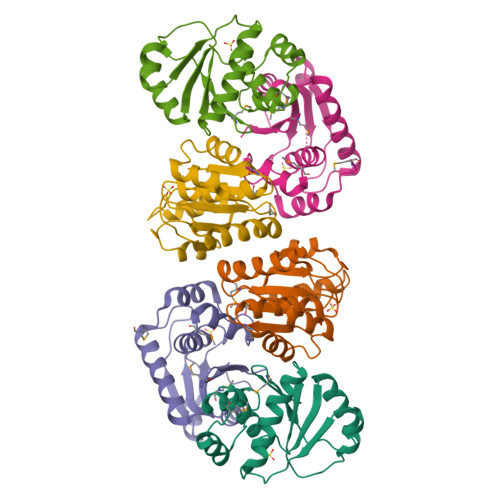
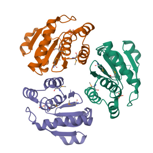
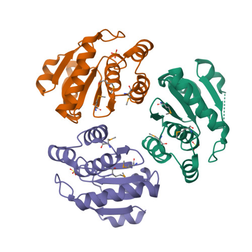
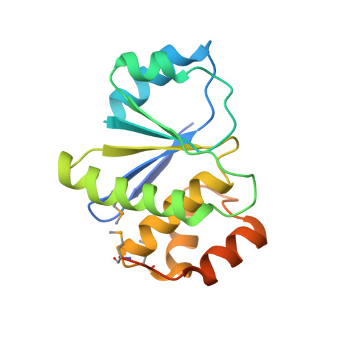
The PRL phosphatases, which constitute a subfamily of the protein tyrosine phosphatases (PTPs), are implicated in oncogenic and metastatic processes. Here, we report the crystal structure of human PRL-1 determined at 2.7A resolution. The crystal structure reveals the shallow active-site pocket with highly hydrophobic character. A structural comparison with the previously determined NMR structure of PRL-3 exhibits significant differences in the active-site region. In the PRL-1 structure, a sulfate ion is bound to the active-site, providing stabilizing interactions to maintain the canonically found active conformation of PTPs, whereas the NMR structure exhibits an open conformation of the active-site. We also found that PRL-1 forms a trimer in the crystal and the trimer exists in the membrane fraction of cells, suggesting the possible biological regulation of PRL-1 activity by oligomerization. The detailed structural information on the active enzyme conformation and regulation of PRL-1 provides the structural basis for the development of potential inhibitors of PRL enzymes.
Organizational Affiliation:
Center for Cellular Switch Protein Structure, Korea Research Institute of Bioscience and Biotechnology, 52 Euh-eun-dong, Yuseong-gu, Daejeon 305-806, South Korea.


