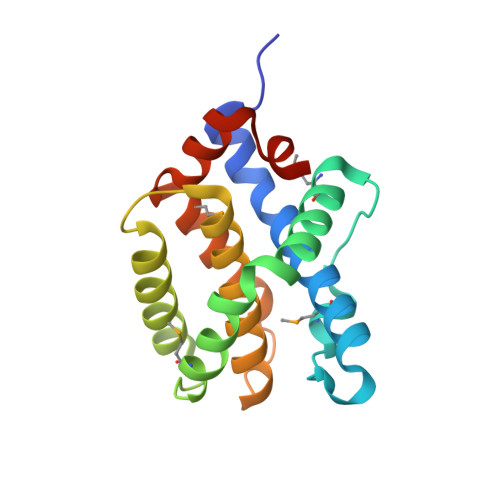
2IMS

The X-ray Structure of a Bak Homodimer Reveals an Inhibitory Zinc Binding Site


Experimental Data Snapshot

  • Method: X-RAY DIFFRACTION
  • Resolution: 1.48 Å
  • R-Value Free: 0.204 
  • R-Value Work: 0.179 
  • R-Value Observed: 0.180 

wwPDB Validation   3D Report Full Report


This is version 1.4 of the entry. See complete history


Literature

The X-Ray Structure of a BAK Homodimer Reveals an Inhibitory Zinc Binding Site

Moldoveanu, T.Liu, Q.Tocilj, A.Watson, M.Shore, G.Gehring, K.

(2006) Mol Cell 24: 677-688

  • DOI: https://doi.org/10.1016/j.molcel.2006.10.014
  • Primary Citation of Related Structures:  
    2IMS, 2IMT

  • PubMed Abstract: 

    BAK/BAX-mediated mitochondrial outer-membrane permeabilization (MOMP) drives cell death during development and tissue homeostasis from zebrafish to humans. In most cancers, this pathway is inhibited by BCL-2 family antiapoptotic members, which bind and block the action of proapoptotic BCL proteins. We report the 1.5 A crystal structure of calpain-proteolysed BAK, cBAK, to reveal a zinc binding site that regulates its activity via homodimerization. cBAK contains an occluded BH3 peptide binding pocket that binds a BID BH3 peptide only weakly . Nonetheless, cBAK requires activation by truncated BID to induce cytochrome c release in mitochondria isolated from bak/bax double-knockout mouse embryonic fibroblasts. The BAK-mediated MOMP is inhibited by low micromolar zinc levels. This inhibition is alleviated by mutation of the zinc-coordination site in BAK. Our results link directly the antiapoptotic effects of zinc to BAK.


  • Organizational Affiliation

    Department of Biochemistry, McGill University, 3655 Promenade Sir William Osler, Montreal, QC, Canada H3G 1Y6. Electronic address: tudor.moldoveanu@stjude.org.


Macromolecules
Find similar proteins by:  (by identity cutoff)  |  3D Structure
Entity ID: 1
MoleculeChains Sequence LengthOrganismDetailsImage
Apoptosis regulator BAK171Homo sapiensMutation(s): 4 
Gene Names: BAK1BAKBCL2L7
UniProt & NIH Common Fund Data Resources
Find proteins for Q16611 (Homo sapiens)
Explore Q16611 
Go to UniProtKB:  Q16611
PHAROS:  Q16611
GTEx:  ENSG00000030110 
Entity Groups  
Sequence Clusters30% Identity50% Identity70% Identity90% Identity95% Identity100% Identity
UniProt GroupQ16611
Sequence Annotations
Expand
  • Reference Sequence
Small Molecules
Ligands 1 Unique
IDChains Name / Formula / InChI Key2D Diagram3D Interactions
ZN
Query on ZN

Download Ideal Coordinates CCD File 
B [auth A]ZINC ION
Zn
PTFCDOFLOPIGGS-UHFFFAOYSA-N
Modified Residues  1 Unique
IDChains TypeFormula2D DiagramParent
MSE
Query on MSE
A
L-PEPTIDE LINKINGC5 H11 N O2 SeMET
Experimental Data & Validation

Experimental Data

  • Method: X-RAY DIFFRACTION
  • Resolution: 1.48 Å
  • R-Value Free: 0.204 
  • R-Value Work: 0.179 
  • R-Value Observed: 0.180 
  • Space Group: C 1 2 1
Unit Cell:
Length ( Å )Angle ( ˚ )
a = 57.527α = 90
b = 53.662β = 115.05
c = 58.281γ = 90
Software Package:
Software NamePurpose
DENZOdata reduction
SCALEPACKdata scaling
REFMACrefinement
PDB_EXTRACTdata extraction
ADSCdata collection
HKL-2000data reduction
SOLVEphasing

Structure Validation

View Full Validation Report


Entry History 

Deposition Data

Revision History  (Full details and data files)

  • Version 1.0: 2006-12-26
    Type: Initial release
  • Version 1.1: 2008-05-01
    Changes: Version format compliance
  • Version 1.2: 2011-07-13
    Changes: Derived calculations, Version format compliance
  • Version 1.3: 2017-10-18
    Changes: Refinement description
  • Version 1.4: 2024-10-16
    Changes: Data collection, Database references, Derived calculations, Structure summary