2ORV

human Thymidine Kinase 1 in complex with TP4A


Experimental Data Snapshot

  • Method: X-RAY DIFFRACTION
  • Resolution: 2.30 Å
  • R-Value Free: 0.249 
  • R-Value Work: 0.197 
  • R-Value Observed: 0.202 

wwPDB Validation   3D Report Full Report


Ligand Structure Quality Assessment 


This is version 1.4 of the entry. See complete history


Literature

Binding of ATP to TK1-like Enzymes Is Associated with a Conformational Change in the Quaternary Structure.

Segura-Pena, D.Lutz, S.Monnerjahn, C.Konrad, M.Lavie, A.

(2007) J Mol Biol 369: 129-141

  • DOI: https://doi.org/10.1016/j.jmb.2007.02.104
  • Primary Citation of Related Structures:  
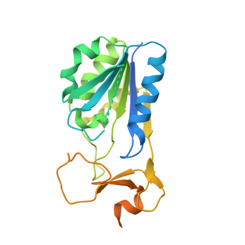
    2ORV, 2ORW

  • PubMed Abstract: 

    Human thymidine kinase 1 (hTK1) and structurally related TKs from other organisms catalyze the initial phosphorylation step in the thymidine salvage pathway. Though ATP is known to be the preferred phosphoryl donor for TK1-like enzymes, its exact binding mode and effect on the oligomeric state has not been analyzed. Here we report the structures of hTK1 and of the Thermotoga maritima thymidine kinase (TmTK) in complex with the bisubstrate inhibitor TP4A. The TmTK-TP4A structure reveals that the adenosine moiety of ATP binds at the subunit interface of the homotetrameric enzyme and that the majority of the ATP-enzyme interactions occur between the phosphate groups and the P-loop. In the hTK1 structure the adenosine group of TP4A exhibited no electron density. This difference between hTK1 and TmTK is rationalized by a difference in the conformation of their quaternary structure. A more open conformation, as seen in the TmTK-TP4A complex structure, is required to provide space for the adenosine moiety. Our analysis supports the formation of an analogous open conformation in hTK1 upon ATP binding.


  • Organizational Affiliation

    Department of Biochemistry and Molecular Genetics, University of Illinois at Chicago, 900 South Ashland Avenue, Chicago, IL 60607, USA.


Macromolecules
Find similar proteins by:  (by identity cutoff)  |  3D Structure
Entity ID: 1
MoleculeChains Sequence LengthOrganismDetailsImage
Thymidine kinase
A, B
234Homo sapiensMutation(s): 2 
Gene Names: TK1
EC: 2.7.1.21
UniProt & NIH Common Fund Data Resources
Find proteins for P04183 (Homo sapiens)
Explore P04183 
Go to UniProtKB:  P04183
PHAROS:  P04183
GTEx:  ENSG00000167900 
Entity Groups  
Sequence Clusters30% Identity50% Identity70% Identity90% Identity95% Identity100% Identity
UniProt GroupP04183
Sequence Annotations
Expand
  • Reference Sequence
Small Molecules
Binding Affinity Annotations 
IDSourceBinding Affinity
4TA Binding MOAD:  2ORV Kd: 29 (nM) from 1 assay(s)
Experimental Data & Validation

Experimental Data

  • Method: X-RAY DIFFRACTION
  • Resolution: 2.30 Å
  • R-Value Free: 0.249 
  • R-Value Work: 0.197 
  • R-Value Observed: 0.202 
  • Space Group: P 31 2 1
Unit Cell:
Length ( Å )Angle ( ˚ )
a = 80.98α = 90
b = 80.98β = 90
c = 156.36γ = 120
Software Package:
Software NamePurpose
XSCALEdata scaling
REFMACrefinement
PDB_EXTRACTdata extraction
XDSdata scaling
XDSdata reduction
SOLVEphasing

Structure Validation

View Full Validation Report


Ligand Structure Quality Assessment 


Entry History 

Deposition Data

Revision History  (Full details and data files)

  • Version 1.0: 2007-03-27
    Type: Initial release
  • Version 1.1: 2008-05-01
    Changes: Version format compliance
  • Version 1.2: 2011-07-13
    Changes: Derived calculations, Version format compliance
  • Version 1.3: 2021-10-20
    Changes: Database references, Derived calculations
  • Version 1.4: 2024-02-21
    Changes: Data collection, Refinement description