2OZO

Autoinhibited intact human ZAP-70


Experimental Data Snapshot

  • Method: X-RAY DIFFRACTION
  • Resolution: 2.60 Å
  • R-Value Free: 0.290 
  • R-Value Work: 0.220 
  • R-Value Observed: 0.260 

wwPDB Validation   3D Report Full Report


Ligand Structure Quality Assessment 


This is version 1.4 of the entry. See complete history


Literature

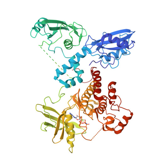
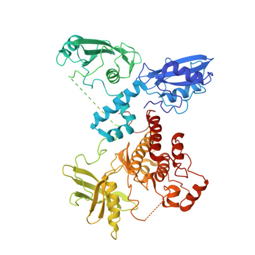
Structural Basis for the Inhibition of Tyrosine Kinase Activity of ZAP-70.

Deindl, S.Kadlecek, T.A.Brdicka, T.Cao, X.Weiss, A.Kuriyan, J.

(2007) Cell 129: 735-746

  • DOI: https://doi.org/10.1016/j.cell.2007.03.039
  • Primary Citation of Related Structures:  
    2OZO

  • PubMed Abstract: 

    ZAP-70, a cytoplasmic tyrosine kinase required for T cell antigen receptor signaling, is controlled by a regulatory segment that includes a tandem SH2 unit responsible for binding to immunoreceptor tyrosine-based activation motifs (ITAMs). The crystal structure of autoinhibited ZAP-70 reveals that the inactive kinase domain adopts a conformation similar to that of cyclin-dependent kinases and Src kinases. The autoinhibitory mechanism of ZAP-70 is, however, distinct and involves interactions between the regulatory segment and the hinge region of the kinase domain that reduce its flexibility. Two tyrosine residues in the SH2-kinase linker that activate ZAP-70 when phosphorylated are involved in aromatic-aromatic interactions that connect the linker to the kinase domain. These interactions are inconsistent with ITAM binding, suggesting that destabilization of this autoinhibited ZAP-70 conformation is the first step in kinase activation.


  • Organizational Affiliation

    Department of Molecular and Cell Biology, Department of Chemistry, and Howard Hughes Medical Institute, University of California, Berkeley, CA 94720, USA.


Macromolecules
Find similar proteins by:  (by identity cutoff)  |  3D Structure
Entity ID: 1
MoleculeChains Sequence LengthOrganismDetailsImage
Tyrosine-protein kinase ZAP-70613Homo sapiensMutation(s): 3 
Gene Names: ZAP70SRK
EC: 2.7.10.2
UniProt & NIH Common Fund Data Resources
Find proteins for P43403 (Homo sapiens)
Explore P43403 
Go to UniProtKB:  P43403
PHAROS:  P43403
GTEx:  ENSG00000115085 
Entity Groups  
Sequence Clusters30% Identity50% Identity70% Identity90% Identity95% Identity100% Identity
UniProt GroupP43403
Sequence Annotations
Expand
  • Reference Sequence
Small Molecules
Ligands 2 Unique
IDChains Name / Formula / InChI Key2D Diagram3D Interactions
ANP
Query on ANP

Download Ideal Coordinates CCD File 
C [auth A]PHOSPHOAMINOPHOSPHONIC ACID-ADENYLATE ESTER
C10 H17 N6 O12 P3
PVKSNHVPLWYQGJ-KQYNXXCUSA-N
MG
Query on MG

Download Ideal Coordinates CCD File 
B [auth A]MAGNESIUM ION
Mg
JLVVSXFLKOJNIY-UHFFFAOYSA-N
Experimental Data & Validation

Experimental Data

  • Method: X-RAY DIFFRACTION
  • Resolution: 2.60 Å
  • R-Value Free: 0.290 
  • R-Value Work: 0.220 
  • R-Value Observed: 0.260 
  • Space Group: P 1
Unit Cell:
Length ( Å )Angle ( ˚ )
a = 48.297α = 105.91
b = 52.929β = 92.94
c = 69.331γ = 103.72
Software Package:
Software NamePurpose
CNSrefinement
PDB_EXTRACTdata extraction

Structure Validation

View Full Validation Report


Ligand Structure Quality Assessment 


Entry History 

Deposition Data

Revision History  (Full details and data files)

  • Version 1.0: 2007-05-22
    Type: Initial release
  • Version 1.1: 2008-05-01
    Changes: Version format compliance
  • Version 1.2: 2011-07-13
    Changes: Version format compliance
  • Version 1.3: 2021-10-20
    Changes: Database references, Derived calculations
  • Version 1.4: 2024-02-21
    Changes: Data collection