2RA3

Human cationic trypsin complexed with bovine pancreatic trypsin inhibitor (BPTI)


Experimental Data Snapshot

  • Method: X-RAY DIFFRACTION
  • Resolution: 1.46 Å
  • R-Value Free: 0.178 
  • R-Value Work: 0.153 
  • R-Value Observed: 0.154 

wwPDB Validation   3D Report Full Report


This is version 1.3 of the entry. See complete history


Literature

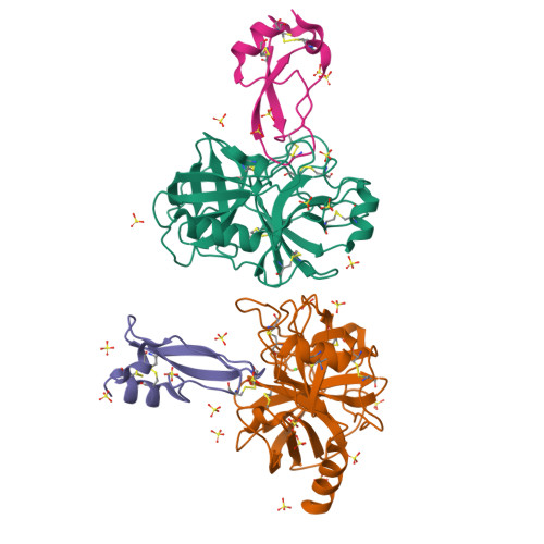
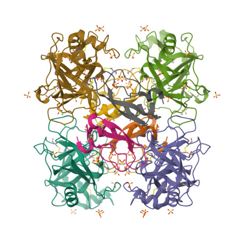
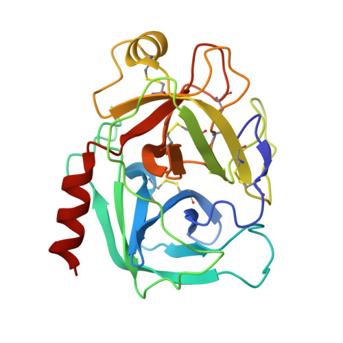
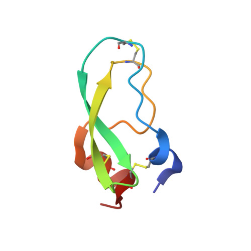
Structural Basis for Accelerated Cleavage of Bovine Pancreatic Trypsin Inhibitor (BPTI) by Human Mesotrypsin.

Salameh, M.A.Soares, A.S.Hockla, A.Radisky, E.S.

(2008) J Biol Chem 283: 4115-4123

  • DOI: https://doi.org/10.1074/jbc.M708268200
  • Primary Citation of Related Structures:  
    2R9P, 2RA3

  • PubMed Abstract: 

    Human mesotrypsin is an isoform of trypsin that displays unusual resistance to polypeptide trypsin inhibitors and has been observed to cleave several such inhibitors as substrates. Whereas substitution of arginine for the highly conserved glycine 193 in the trypsin active site has been implicated as a critical factor in the inhibitor resistance of mesotrypsin, how this substitution leads to accelerated inhibitor cleavage is not clear. Bovine pancreatic trypsin inhibitor (BPTI) forms an extremely stable and cleavage-resistant complex with trypsin, and thus provides a rigorous challenge of mesotrypsin catalytic activity toward polypeptide inhibitors. Here, we report kinetic constants for mesotrypsin and the highly homologous (but inhibitor sensitive) human cationic trypsin, describing inhibition by, and cleavage of BPTI, as well as crystal structures of the mesotrypsin-BPTI and human cationic trypsin-BPTI complexes. We find that mesotrypsin cleaves BPTI with a rate constant accelerated 350-fold over that of human cationic trypsin and 150,000-fold over that of bovine trypsin. From the crystal structures, we see that small conformational adjustments limited to several side chains enable mesotrypsin-BPTI complex formation, surmounting the predicted steric clash introduced by Arg-193. Our results show that the mesotrypsin-BPTI interface favors catalysis through (a) electrostatic repulsion between the closely spaced mesotrypsin Arg-193 and BPTI Arg-17, and (b) elimination of two hydrogen bonds between the enzyme and the amine leaving group portion of BPTI. Our model predicts that these deleterious interactions accelerate leaving group dissociation and deacylation.


  • Organizational Affiliation

    Department of Cancer Biology, Mayo Clinic Cancer Center, Jacksonville, Florida 32224, USA.


Macromolecules
Find similar proteins by:  (by identity cutoff)  |  3D Structure
Entity ID: 1
MoleculeChains Sequence LengthOrganismDetailsImage
Trypsin-1
A, B
224Homo sapiensMutation(s): 2 
Gene Names: PRSS1TRP1TRY1TRYP1
EC: 3.4.21.4
UniProt & NIH Common Fund Data Resources
Find proteins for P07477 (Homo sapiens)
Explore P07477 
Go to UniProtKB:  P07477
PHAROS:  P07477
GTEx:  ENSG00000204983 
Entity Groups  
Sequence Clusters30% Identity50% Identity70% Identity90% Identity95% Identity100% Identity
UniProt GroupP07477
Sequence Annotations
Expand
  • Reference Sequence
Find similar proteins by:  (by identity cutoff)  |  3D Structure
Entity ID: 2
MoleculeChains Sequence LengthOrganismDetailsImage
Pancreatic trypsin inhibitorC [auth I],
D [auth C]
58Bos taurusMutation(s): 0 
UniProt
Find proteins for P00974 (Bos taurus)
Explore P00974 
Go to UniProtKB:  P00974
Entity Groups  
Sequence Clusters30% Identity50% Identity70% Identity90% Identity95% Identity100% Identity
UniProt GroupP00974
Sequence Annotations
Expand
  • Reference Sequence
Small Molecules
Ligands 2 Unique
IDChains Name / Formula / InChI Key2D Diagram3D Interactions
SO4
Query on SO4

Download Ideal Coordinates CCD File 
AA [auth I]
BA [auth I]
CA [auth C]
DA [auth C]
F [auth A]
AA [auth I],
BA [auth I],
CA [auth C],
DA [auth C],
F [auth A],
G [auth A],
H [auth A],
I [auth A],
J [auth A],
K [auth A],
L [auth A],
N [auth B],
O [auth B],
P [auth B],
Q [auth B],
R [auth B],
S [auth B],
T [auth B],
U [auth B],
V [auth B],
W [auth B],
X [auth I],
Y [auth I],
Z [auth I]
SULFATE ION
O4 S
QAOWNCQODCNURD-UHFFFAOYSA-L
CA
Query on CA

Download Ideal Coordinates CCD File 
E [auth A],
M [auth B]
CALCIUM ION
Ca
BHPQYMZQTOCNFJ-UHFFFAOYSA-N
Experimental Data & Validation

Experimental Data

  • Method: X-RAY DIFFRACTION
  • Resolution: 1.46 Å
  • R-Value Free: 0.178 
  • R-Value Work: 0.153 
  • R-Value Observed: 0.154 
  • Space Group: C 2 2 21
Unit Cell:
Length ( Å )Angle ( ˚ )
a = 91.183α = 90
b = 186.546β = 90
c = 90.187γ = 90
Software Package:
Software NamePurpose
ADSCdata collection
PHENIXmodel building
PHENIXrefinement
MOSFLMdata reduction
SCALAdata scaling
PHENIXphasing

Structure Validation

View Full Validation Report


Entry History 

Deposition Data

Revision History  (Full details and data files)

  • Version 1.0: 2007-12-11
    Type: Initial release
  • Version 1.1: 2011-07-13
    Changes: Version format compliance
  • Version 1.2: 2021-10-20
    Changes: Database references, Derived calculations
  • Version 1.3: 2023-08-30
    Changes: Data collection, Refinement description