2UZF

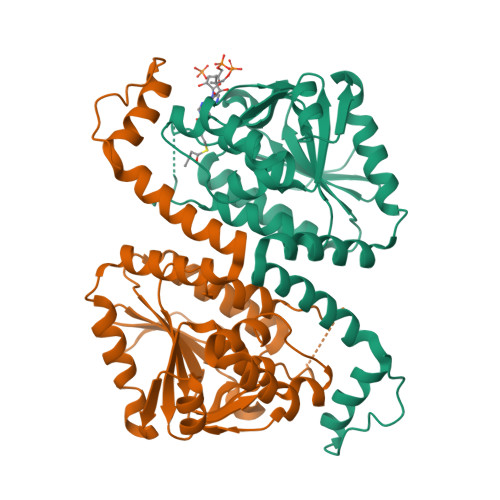
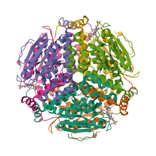
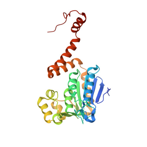
Crystal structure of Staphylococcus aureus 1,4-dihydroxy-2-naphthoyl CoA synthase (MenB) in complex with acetoacetyl CoA


Experimental Data Snapshot

  • Method: X-RAY DIFFRACTION
  • Resolution: 2.90 Å
  • R-Value Free: 0.328 
  • R-Value Work: 0.247 
  • R-Value Observed: 0.250 

Starting Model: experimental
View more details

wwPDB Validation   3D Report Full Report


Ligand Structure Quality Assessment 


This is version 1.3 of the entry. See complete history


Literature

Structure of Staphylococcus Aureus1,4-Dihydroxy-2-Naphthoyl-Coa Synthase (Menb) in Complex with Acetoacetyl-Coa.

Ulaganathan, V.Agacan, M.F.Buetow, L.Tulloch, L.B.Hunter, W.N.

(2007) Acta Crystallogr Sect F Struct Biol Cryst Commun 63: 908

  • DOI: https://doi.org/10.1107/S1744309107047720
  • Primary Citation of Related Structures:  
    2UZF

  • PubMed Abstract: 

    Vitamin K(2), or menaquinone, is an essential cofactor for many organisms and the enzymes involved in its biosynthesis are potential antimicrobial drug targets. One of these enzymes, 1,4-dihydroxy-2-naphthoyl-CoA synthase (MenB) from the pathogen Staphylococcus aureus, has been obtained in recombinant form and its quaternary structure has been analyzed in solution. Cubic crystals of the enzyme allowed a low-resolution structure (2.9 A) to be determined. The asymmetric unit consists of two subunits and a crystallographic threefold axis of symmetry generates a hexamer consistent with size-exclusion chromatography. Analytical ultracentrifugation indicates the presence of six states in solution, monomeric through to hexameric, with the dimer noted as being particularly stable. MenB displays the crotonase-family fold with distinct N- and C-terminal domains and a flexible segment of structure around the active site. The smaller C-terminal domain plays an important role in oligomerization and also in substrate binding. The presence of acetoacetyl-CoA in one of the two active sites present in the asymmetric unit indicates how part of the substrate binds and facilitates comparisons with the structure of Mycobacterium tuberculosis MenB.


  • Organizational Affiliation

    Division of Biological Chemistry and Drug Discovery, College of Life Sciences, University of Dundee, Dundee DD1 5EH, Tayside, Scotland.


Macromolecules
Find similar proteins by:  (by identity cutoff)  |  3D Structure
Entity ID: 1
MoleculeChains Sequence LengthOrganismDetailsImage
NAPHTHOATE SYNTHASE
A, B
273Staphylococcus aureusMutation(s): 0 
EC: 4.1.3.36
UniProt
Find proteins for Q5HH38 (Staphylococcus aureus (strain COL))
Explore Q5HH38 
Go to UniProtKB:  Q5HH38
Entity Groups  
Sequence Clusters30% Identity50% Identity70% Identity90% Identity95% Identity100% Identity
UniProt GroupQ5HH38
Sequence Annotations
Expand
  • Reference Sequence
Small Molecules
Ligands 1 Unique
IDChains Name / Formula / InChI Key2D Diagram3D Interactions
CAA
Query on CAA

Download Ideal Coordinates CCD File 
C [auth A]ACETOACETYL-COENZYME A
C25 H40 N7 O18 P3 S
OJFDKHTZOUZBOS-CITAKDKDSA-N
Experimental Data & Validation

Experimental Data

  • Method: X-RAY DIFFRACTION
  • Resolution: 2.90 Å
  • R-Value Free: 0.328 
  • R-Value Work: 0.247 
  • R-Value Observed: 0.250 
  • Space Group: P 21 3
Unit Cell:
Length ( Å )Angle ( ˚ )
a = 120.008α = 90
b = 120.008β = 90
c = 120.008γ = 90
Software Package:
Software NamePurpose
REFMACrefinement
MOSFLMdata reduction
SCALAdata scaling
PHASERphasing

Structure Validation

View Full Validation Report


Ligand Structure Quality Assessment 


Entry History 

Deposition Data

Revision History  (Full details and data files)

  • Version 1.0: 2007-11-13
    Type: Initial release
  • Version 1.1: 2011-07-13
    Changes: Advisory, Version format compliance
  • Version 1.2: 2012-06-13
    Changes: Other
  • Version 1.3: 2023-12-13
    Changes: Data collection, Database references, Derived calculations, Other, Refinement description