The Crystal Structure of Caspase-6, a Selective Effector of Axonal Degeneration.
Baumgartner, R., Meder, G., Briand, C., Decock, A., D'Arcy, A., Hassiepen, U., Morse, R., Renatus, M.(2009) Biochem J 423: 429
- PubMed: 19694615
- DOI: https://doi.org/10.1042/BJ20090540
- Primary Citation of Related Structures:
2WDP - PubMed Abstract:
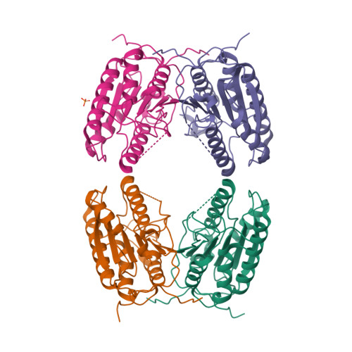
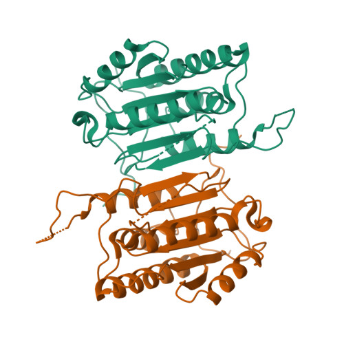
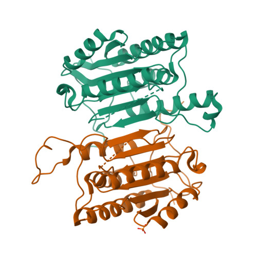
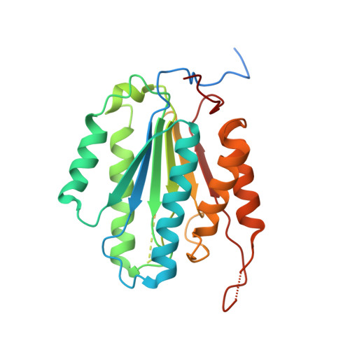
Neurodegenerative diseases pose one of the most pressing unmet medical needs today. It has long been recognized that caspase-6 may play a role in several neurodegenerative diseases for which there are currently no disease-modifying therapies. Thus it is a potential target for neurodegenerative drug development. In the present study we report on the biochemistry and structure of caspase-6. As an effector caspase, caspase-6 is a constitutive dimer independent of the maturation state of the enzyme. The ligand-free structure shows caspase-6 in a partially mature but latent conformation. The cleaved inter-domain linker remains partially inserted in the central groove of the dimer, as observed in other caspases. However, in contrast with the structures of other caspases, not only is the catalytic machinery misaligned, but several structural elements required for substrate recognition are missing. Most importantly, residues forming a short anti-parallel beta-sheet abutting the substrate in other caspase structures are part of an elongation of the central alpha-helix. Despite the dramatic structural changes that are required to adopt a canonical catalytically competent conformation, the pre-steady-state kinetics exhibit no lag phase in substrate turnover. This suggests that the observed conformation does not play a regulatory role in caspase-6 activity. However, targeting the latent conformation in search for specific and bio-available caspase-6 inhibitors might offer an alternative to active-site-directed approaches.
Organizational Affiliation:
Novartis Institutes for BioMedical Research, Center for Proteomic Chemistry, Expertise Platform Proteases, 4002 Basel, Switzerland.


