3B4V | pdb_00003b4v

X-Ray structure of Activin in complex with FSTL3


Experimental Data Snapshot

  • Method: X-RAY DIFFRACTION
  • Resolution: 2.48 Å
  • R-Value Free: 
    0.279 (Depositor), 0.266 (DCC) 
  • R-Value Work: 
    0.227 (Depositor), 0.242 (DCC) 
  • R-Value Observed: 
    0.229 (Depositor) 

wwPDB Validation   3D Report Full Report


Ligand Structure Quality Assessment 

Created with Raphaël 2.3.0Worse 01 BetterLigand structure goodness of fit to experimental dataBest fitted NAGClick on this verticalbar to view details

This is version 1.4 of the entry. See complete history


Literature

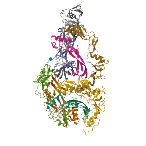
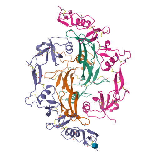
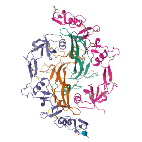
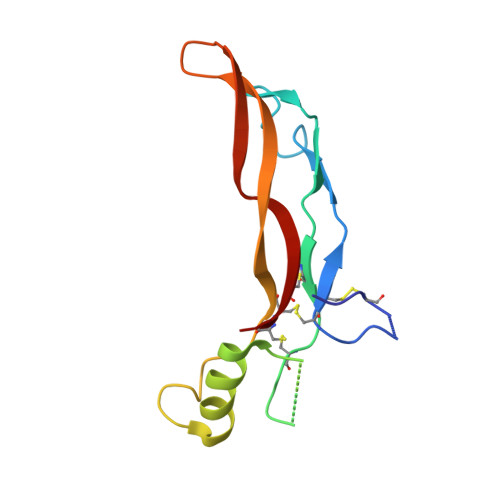
The structure of FSTL3.activin A complex. Differential binding of N-terminal domains influences follistatin-type antagonist specificity.

Stamler, R.Keutmann, H.T.Sidis, Y.Kattamuri, C.Schneyer, A.Thompson, T.B.

(2008) J Biological Chem 283: 32831-32838

  • DOI: https://doi.org/10.1074/jbc.M801266200
  • Primary Citation of Related Structures:  
    3B4V

  • PubMed Abstract: 

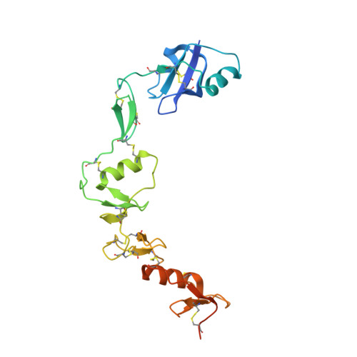
    Transforming growth factor beta family ligands are neutralized by a number of structurally divergent antagonists. Follistatin-type antagonists, which include splice variants of follistatin (FS288 and FS315) and follistatin-like 3 (FSTL3), have high affinity for activin A but differ in their affinity for other ligands, particularly bone morphogenetic proteins. To understand the structural basis for ligand specificity within FS-type antagonists, we determined the x-ray structure of activin A in complex with FSTL3 to a resolution of 2.5 A. Similar to the previously resolved FS.activin A structures, the ligand is encircled by two antagonist molecules blocking all ligand receptor-binding sites. Recently, the significance of the FS N-terminal domain interaction at the ligand type I receptor site has been questioned; however, our data show that for FSTL3, the N-terminal domain forms a more intimate contact with activin A, implying that this interaction is stronger than that for FS. Furthermore, binding studies revealed that replacing the FSTL3 N-terminal domain with the corresponding FS domain considerably lowers activin A affinity. Therefore, both structural and biochemical evidence support a significant interaction of the N-terminal domain of FSTL3 with activin A. In addition, structural comparisons with bone morphogenetic proteins suggest that the interface where the N-terminal domain binds may be the key site for determining FS-type antagonist specificity.


  • Organizational Affiliation

    Department of Molecular Genetics, Biochemistry and Microbiology, University of Cincinnati Medical Sciences Building, Cincinnati, Ohio 45267, USA.


Macromolecules
Find similar proteins by:  (by identity cutoff)  |  3D Structure
Entity ID: 1
MoleculeChains Sequence LengthOrganismDetailsImage
Inhibin beta A chain
A, B, E, F
116Homo sapiensMutation(s): 0 
Gene Names: INHBA
UniProt & NIH Common Fund Data Resources
Find proteins for P08476 (Homo sapiens)
Explore P08476 
Go to UniProtKB:  P08476
PHAROS:  P08476
GTEx:  ENSG00000122641 
Entity Groups  
Sequence Clusters30% Identity50% Identity70% Identity90% Identity95% Identity100% Identity
UniProt GroupP08476
Sequence Annotations
Expand
  • Reference Sequence
Find similar proteins by:  (by identity cutoff)  |  3D Structure
Entity ID: 2
MoleculeChains Sequence LengthOrganismDetailsImage
follistatin-like 3
C, D, G, H
237Homo sapiensMutation(s): 0 
Gene Names: FSTL3FLRG
UniProt & NIH Common Fund Data Resources
Find proteins for O95633 (Homo sapiens)
Explore O95633 
Go to UniProtKB:  O95633
PHAROS:  O95633
GTEx:  ENSG00000070404 
Entity Groups  
Sequence Clusters30% Identity50% Identity70% Identity90% Identity95% Identity100% Identity
UniProt GroupO95633
Glycosylation
Glycosylation Sites: 1Go to GlyGen: O95633-1
Sequence Annotations
Expand
  • Reference Sequence
Experimental Data & Validation

Experimental Data

  • Method: X-RAY DIFFRACTION
  • Resolution: 2.48 Å
  • R-Value Free:  0.279 (Depositor), 0.266 (DCC) 
  • R-Value Work:  0.227 (Depositor), 0.242 (DCC) 
  • R-Value Observed: 0.229 (Depositor) 
Space Group: P 1
Unit Cell:
Length ( Å )Angle ( ˚ )
a = 63.613α = 98.55
b = 71.384β = 90.64
c = 100.203γ = 90.11
Software Package:
Software NamePurpose
DENZOdata reduction
SCALEPACKdata scaling
PHASERphasing
REFMACrefinement
PDB_EXTRACTdata extraction
HKL-2000data collection
HKL-2000data reduction
HKL-2000data scaling

Structure Validation

View Full Validation Report


Ligand Structure Quality Assessment 

Created with Raphaël 2.3.0Worse 01 BetterLigand structure goodness of fit to experimental dataBest fitted NAGClick on this verticalbar to view details

Entry History 

Deposition Data

Revision History  (Full details and data files)

  • Version 1.0: 2008-09-02
    Type: Initial release
  • Version 1.1: 2011-07-13
    Changes: Non-polymer description, Version format compliance
  • Version 1.2: 2017-10-25
    Changes: Refinement description
  • Version 1.3: 2020-07-29
    Type: Remediation
    Reason: Carbohydrate remediation
    Changes: Data collection, Derived calculations, Structure summary
  • Version 1.4: 2024-10-30
    Changes: Data collection, Database references, Structure summary