3C5V

PP2A-specific methylesterase apo form (PME)


Experimental Data Snapshot

  • Method: X-RAY DIFFRACTION
  • Resolution: 2.00 Å
  • R-Value Free: 0.220 
  • R-Value Work: 0.183 
  • R-Value Observed: 0.183 

wwPDB Validation   3D Report Full Report


This is version 1.3 of the entry. See complete history


Literature

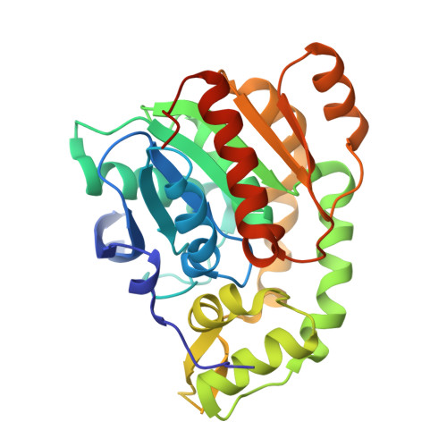
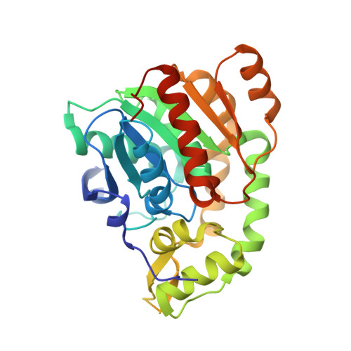
Structural mechanism of demethylation and inactivation of protein phosphatase 2A.

Xing, Y.Li, Z.Chen, Y.Stock, J.B.Jeffrey, P.D.Shi, Y.

(2008) Cell 133: 154-163

  • DOI: https://doi.org/10.1016/j.cell.2008.02.041
  • Primary Citation of Related Structures:  
    3C5V, 3C5W

  • PubMed Abstract: 

    Protein phosphatase 2A (PP2A) is an important serine/threonine phosphatase that plays a role in many biological processes. Reversible carboxyl methylation of the PP2A catalytic subunit is an essential regulatory mechanism for its function. Demethylation and negative regulation of PP2A is mediated by a PP2A-specific methylesterase PME-1, which is conserved from yeast to humans. However, the underlying mechanism of PME-1 function remains enigmatic. Here we report the crystal structures of PME-1 by itself and in complex with a PP2A heterodimeric core enzyme. The structures reveal that PME-1 directly binds to the active site of PP2A and that this interaction results in the activation of PME-1 by rearranging the catalytic triad into an active conformation. Strikingly, these interactions also lead to inactivation of PP2A by evicting the manganese ions that are required for the phosphatase activity of PP2A. These observations identify a dual role of PME-1 that regulates PP2A activation, methylation, and holoenzyme assembly in cells.


  • Organizational Affiliation

    Department of Molecular Biology, Princeton University, Lewis Thomas Laboratory, Princeton, NJ 08544, USA.


Macromolecules
Find similar proteins by:  (by identity cutoff)  |  3D Structure
Entity ID: 1
MoleculeChains Sequence LengthOrganismDetailsImage
Protein phosphatase methylesterase 1316Homo sapiensMutation(s): 0 
Gene Names: PPME1PME1
UniProt & NIH Common Fund Data Resources
Find proteins for Q9Y570 (Homo sapiens)
Explore Q9Y570 
Go to UniProtKB:  Q9Y570
PHAROS:  Q9Y570
GTEx:  ENSG00000214517 
Entity Groups  
Sequence Clusters30% Identity50% Identity70% Identity90% Identity95% Identity100% Identity
UniProt GroupQ9Y570
Sequence Annotations
Expand
  • Reference Sequence
Experimental Data & Validation

Experimental Data

  • Method: X-RAY DIFFRACTION
  • Resolution: 2.00 Å
  • R-Value Free: 0.220 
  • R-Value Work: 0.183 
  • R-Value Observed: 0.183 
  • Space Group: P 31 2 1
Unit Cell:
Length ( Å )Angle ( ˚ )
a = 82.512α = 90
b = 82.512β = 90
c = 90.843γ = 120
Software Package:
Software NamePurpose
CNSrefinement
CBASSdata collection
DENZOdata reduction
SCALEPACKdata scaling
SHELXSphasing

Structure Validation

View Full Validation Report


Entry History 

Deposition Data

Revision History  (Full details and data files)

  • Version 1.0: 2008-04-15
    Type: Initial release
  • Version 1.1: 2011-07-13
    Changes: Version format compliance
  • Version 1.2: 2017-07-26
    Changes: Source and taxonomy
  • Version 1.3: 2024-02-21
    Changes: Data collection, Database references