3DI2

Crystal structure of the complex of human interleukin-7 with unglycosylated human interleukin-7 receptor alpha ectodomain


Experimental Data Snapshot

  • Method: X-RAY DIFFRACTION
  • Resolution: 2.70 Å
  • R-Value Free: 0.267 
  • R-Value Work: 0.212 
  • R-Value Observed: 0.212 

wwPDB Validation   3D Report Full Report


This is version 1.2 of the entry. See complete history


Literature

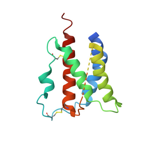
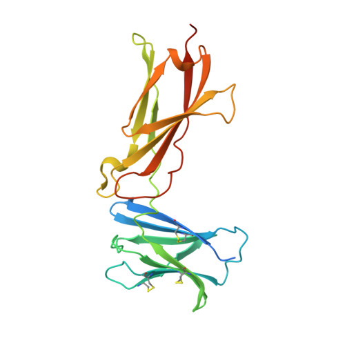
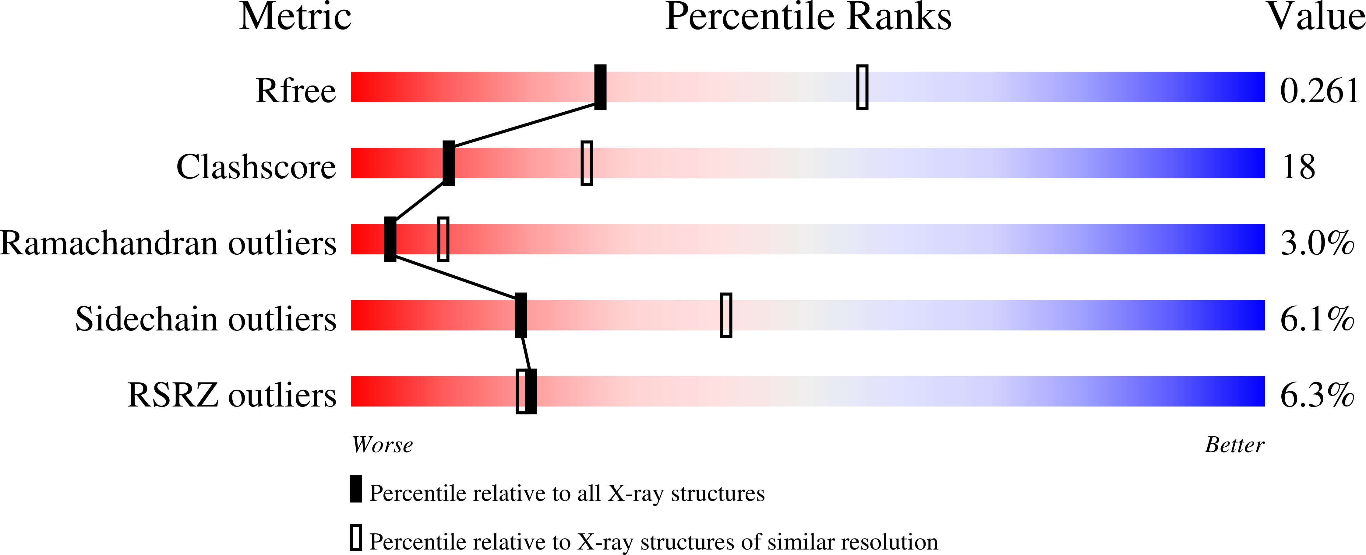
Structural and Biophysical Studies of the Human IL-7/IL-7Ralpha Complex.

McElroy, C.A.Dohm, J.A.Walsh, S.T.

(2009) Structure 17: 54-65

  • DOI: https://doi.org/10.1016/j.str.2008.10.019
  • Primary Citation of Related Structures:  
    3DI2, 3DI3

  • PubMed Abstract: 

    IL-7 and IL-7Ralpha bind the gamma(c) receptor, forming a complex crucial to several signaling cascades leading to the development and homeostasis of T and B cells. We report that the IL-7Ralpha ectodomain uses glycosylation to modulate its binding constants to IL-7, unlike the other receptors in the gamma(c) family. IL-7 binds glycosylated IL-7Ralpha 300-fold more tightly than unglycosylated IL-7Ralpha, and the enhanced affinity is attributed primarily to an accelerated on rate. Structural comparison of IL-7 in complex to both forms of IL-7Ralpha reveals that glycosylation does not participate directly in the binding interface. The SCID mutations of IL-7Ralpha locate outside the binding interface with IL-7, suggesting that the expressed mutations cause protein folding defects in IL-7Ralpha. The IL-7/IL-7Ralpha structures provide a window into the molecular recognition events of the IL-7 signaling cascade and provide sites to target for designing new therapeutics to treat IL-7-related diseases.


  • Organizational Affiliation

    Department of Molecular and Cellular Biochemistry, College of Medicine, Comprehensive Cancer Center, Ohio State University, 467 Hamilton Hall, 1645 Neil Avenue, Columbus, OH 43210, USA.


Macromolecules
Find similar proteins by:  (by identity cutoff)  |  3D Structure
Entity ID: 1
MoleculeChains Sequence LengthOrganismDetailsImage
Interleukin-7
A, C
154Homo sapiensMutation(s): 1 
Gene Names: IL7
UniProt & NIH Common Fund Data Resources
Find proteins for P13232 (Homo sapiens)
Explore P13232 
Go to UniProtKB:  P13232
PHAROS:  P13232
GTEx:  ENSG00000104432 
Entity Groups  
Sequence Clusters30% Identity50% Identity70% Identity90% Identity95% Identity100% Identity
UniProt GroupP13232
Sequence Annotations
Expand
  • Reference Sequence
Find similar proteins by:  (by identity cutoff)  |  3D Structure
Entity ID: 2
MoleculeChains Sequence LengthOrganismDetailsImage
Interleukin-7 receptor subunit alpha
B, D
223Homo sapiensMutation(s): 1 
Gene Names: IL7R
UniProt & NIH Common Fund Data Resources
Find proteins for P16871 (Homo sapiens)
Explore P16871 
Go to UniProtKB:  P16871
PHAROS:  P16871
GTEx:  ENSG00000168685 
Entity Groups  
Sequence Clusters30% Identity50% Identity70% Identity90% Identity95% Identity100% Identity
UniProt GroupP16871
Sequence Annotations
Expand
  • Reference Sequence
Small Molecules
Ligands 1 Unique
IDChains Name / Formula / InChI Key2D Diagram3D Interactions
1PE
Query on 1PE

Download Ideal Coordinates CCD File 
E [auth A]PENTAETHYLENE GLYCOL
C10 H22 O6
JLFNLZLINWHATN-UHFFFAOYSA-N
Experimental Data & Validation

Experimental Data

  • Method: X-RAY DIFFRACTION
  • Resolution: 2.70 Å
  • R-Value Free: 0.267 
  • R-Value Work: 0.212 
  • R-Value Observed: 0.212 
  • Space Group: P 1 21 1
Unit Cell:
Length ( Å )Angle ( ˚ )
a = 47.202α = 90
b = 113.021β = 105.1
c = 87.534γ = 90
Software Package:
Software NamePurpose
CNSrefinement
PDB_EXTRACTdata extraction
HKL-2000data collection
HKL-2000data reduction
HKL-2000data scaling

Structure Validation

View Full Validation Report


Entry History 

Deposition Data

Revision History  (Full details and data files)

  • Version 1.0: 2009-01-27
    Type: Initial release
  • Version 1.1: 2011-07-13
    Changes: Version format compliance
  • Version 1.2: 2021-10-20
    Changes: Database references, Derived calculations