3G33

Crystal structure of CDK4/cyclin D3


Experimental Data Snapshot

  • Method: X-RAY DIFFRACTION
  • Resolution: 3.00 Å
  • R-Value Free: 0.314 
  • R-Value Work: 0.281 
  • R-Value Observed: 0.283 

wwPDB Validation   3D Report Full Report


This is version 1.3 of the entry. See complete history


Literature

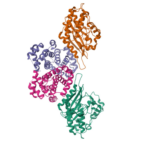
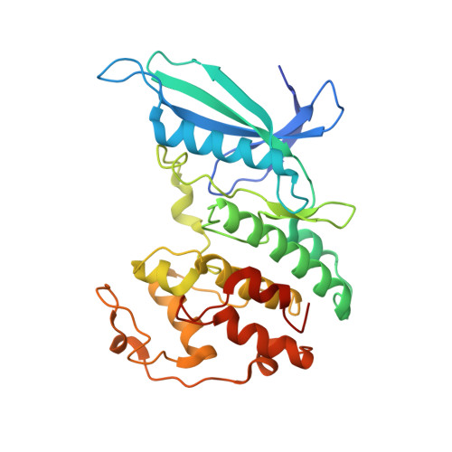
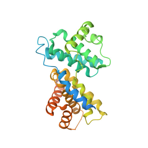
The structure of CDK4/cyclin D3 has implications for models of CDK activation.

Takaki, T.Echalier, A.Brown, N.R.Hunt, T.Endicott, J.A.Noble, M.E.

(2009) Proc Natl Acad Sci U S A 106: 4171-4176

  • DOI: https://doi.org/10.1073/pnas.0809674106
  • Primary Citation of Related Structures:  
    3G33

  • PubMed Abstract: 

    Cyclin-dependent kinase 4 (CDK4)/cyclin D complexes are expressed early in the G(1) phase of the cell cycle and stimulate the expression of genes required for G(1) progression by phosphorylation of the product of the retinoblastoma gene, pRb. To elaborate the molecular pathway of CDK4 activation and substrate selection we have determined the structure of nonphosphorylated CDK4/cyclin D3. This structure of an authentic CDK/cyclin complex shows that cyclin binding may not be sufficient to drive the CDK active site toward an active conformation. Phosphorylated CDK4/cyclin D3 is active as a pRb kinase and is susceptible to inhibition by p27(Kip1). Unlike CDK2/cyclin A, CDK4/cyclin D3 can be inactivated by treatment with lambda-phosphatase, implying that phosphorylated T172 is accessible to a generic phosphatase while bound to a cyclin. Taken together, these results suggest that the structural mechanism of CDK4/cyclin D3 activation differs markedly from that of previously studied CDK/cyclin complexes.


  • Organizational Affiliation

    Cell Cycle Control Laboratory, Clare Hall Laboratories, London Research Institute, Blanche Lane, South Mimms, Herts EN6 3LD, United Kingdom.


Macromolecules
Find similar proteins by:  (by identity cutoff)  |  3D Structure
Entity ID: 1
MoleculeChains Sequence LengthOrganismDetailsImage
Cell division protein kinase 4
A, C
308Homo sapiensMutation(s): 0 
Gene Names: CDK4
EC: 2.7.11.22
UniProt & NIH Common Fund Data Resources
Find proteins for P11802 (Homo sapiens)
Explore P11802 
Go to UniProtKB:  P11802
PHAROS:  P11802
GTEx:  ENSG00000135446 
Entity Groups  
Sequence Clusters30% Identity50% Identity70% Identity90% Identity95% Identity100% Identity
UniProt GroupP11802
Sequence Annotations
Expand
  • Reference Sequence
Find similar proteins by:  (by identity cutoff)  |  3D Structure
Entity ID: 2
MoleculeChains Sequence LengthOrganismDetailsImage
CCND3 protein
B, D
306Homo sapiensMutation(s): 0 
Gene Names: CCND3hCG_16683
UniProt & NIH Common Fund Data Resources
Find proteins for P30281 (Homo sapiens)
Explore P30281 
Go to UniProtKB:  P30281
PHAROS:  P30281
GTEx:  ENSG00000112576 
Entity Groups  
Sequence Clusters30% Identity50% Identity70% Identity90% Identity95% Identity100% Identity
UniProt GroupP30281
Sequence Annotations
Expand
  • Reference Sequence
Experimental Data & Validation

Experimental Data

  • Method: X-RAY DIFFRACTION
  • Resolution: 3.00 Å
  • R-Value Free: 0.314 
  • R-Value Work: 0.281 
  • R-Value Observed: 0.283 
  • Space Group: P 42 21 2
Unit Cell:
Length ( Å )Angle ( ˚ )
a = 141.297α = 90
b = 141.297β = 90
c = 143.53γ = 90
Software Package:
Software NamePurpose
ADSCdata collection
PHASERphasing
REFMACrefinement
MOSFLMdata reduction
SCALEPACKdata scaling

Structure Validation

View Full Validation Report


Entry History 

Deposition Data

Revision History  (Full details and data files)

  • Version 1.0: 2009-03-10
    Type: Initial release
  • Version 1.1: 2011-07-13
    Changes: Advisory, Version format compliance
  • Version 1.2: 2017-08-23
    Changes: Data collection
  • Version 1.3: 2024-02-21
    Changes: Data collection, Database references