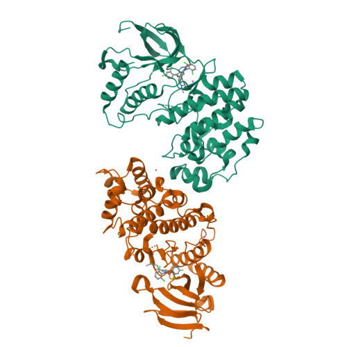
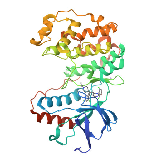
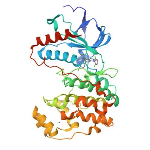
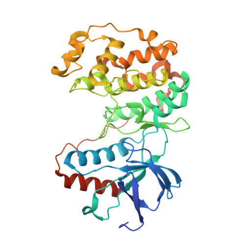
3GC9

The structure of p38beta C119S, C162S in complex with a dihydroquinazolinone inhibitor


Experimental Data Snapshot

  • Method: X-RAY DIFFRACTION
  • Resolution: 2.05 Å
  • R-Value Free: 0.275 
  • R-Value Work: 0.222 
  • R-Value Observed: 0.224 

Starting Model: experimental
View more details

wwPDB Validation   3D Report Full Report


Ligand Structure Quality Assessment 


This is version 1.5 of the entry. See complete history


Literature

The three-dimensional structure of MAP kinase p38beta: different features of the ATP-binding site in p38beta compared with p38alpha.

Patel, S.B.Cameron, P.M.O'Keefe, S.J.Frantz-Wattley, B.Thompson, J.O'Neill, E.A.Tennis, T.Liu, L.Becker, J.W.Scapin, G.

(2009) Acta Crystallogr D Biol Crystallogr 65: 777-785

  • DOI: https://doi.org/10.1107/S090744490901600X
  • Primary Citation of Related Structures:  
    3GC7, 3GC8, 3GC9

  • PubMed Abstract: 

    The p38 mitogen-activated protein kinases are activated in response to environmental stress and cytokines and play a significant role in transcriptional regulation and inflammatory responses. Of the four p38 isoforms known to date, two (p38alpha and p38beta) have been identified as targets for cytokine-suppressive anti-inflammatory drugs. Recently, it was reported that specific inhibition of the p38alpha isoform is necessary and sufficient for anti-inflammatory efficacy in vivo, while further inhibition of p38beta may not provide any additional benefit. In order to aid the development of p38alpha-selective compounds, the three-dimensional structure of p38beta was determined. To do so, the C162S and C119S,C162S mutants of human MAP kinase p38beta were cloned, expressed in Escherichia coli and purified. Initial screening hits in crystallization trials in the presence of an inhibitor led upon optimization to crystals that diffracted to 2.05 A resolution and allowed structure determination (PDB codes 3gc8 and 3gc9 for the single and double mutant, respectively). The structure of the p38alpha C162S mutant in complex with the same inhibitor is also reported (PDB code 3gc7). A comparison between the structures of the two kinases showed that they are highly similar overall but that there are differences in the relative orientation of the N- and C-terminal domains that causes a reduction in the size of the ATP-binding pocket in p38beta. This difference in size between the two pockets could be exploited in order to achieve selectivity.


  • Organizational Affiliation

    Department of Global Structural Biology, Merck Research Laboratory, Rahway, NJ 07065, USA.


Macromolecules
Find similar proteins by:  (by identity cutoff)  |  3D Structure
Entity ID: 1
MoleculeChains Sequence LengthOrganismDetailsImage
Mitogen-activated protein kinase 11
A, B
370Homo sapiensMutation(s): 2 
Gene Names: MAPK11PRKM11SAPK2
EC: 2.7.11.24
UniProt & NIH Common Fund Data Resources
Find proteins for Q15759 (Homo sapiens)
Explore Q15759 
Go to UniProtKB:  Q15759
PHAROS:  Q15759
GTEx:  ENSG00000185386 
Entity Groups  
Sequence Clusters30% Identity50% Identity70% Identity90% Identity95% Identity100% Identity
UniProt GroupQ15759
Sequence Annotations
Expand
  • Reference Sequence
Small Molecules
Binding Affinity Annotations 
IDSourceBinding Affinity
B45 BindingDB:  3GC9 IC50: min: 0.6, max: 5.6 (nM) from 2 assay(s)
Experimental Data & Validation

Experimental Data

  • Method: X-RAY DIFFRACTION
  • Resolution: 2.05 Å
  • R-Value Free: 0.275 
  • R-Value Work: 0.222 
  • R-Value Observed: 0.224 
  • Space Group: P 1 21 1
Unit Cell:
Length ( Å )Angle ( ˚ )
a = 39.184α = 90
b = 158.823β = 91.59
c = 60.874γ = 90
Software Package:
Software NamePurpose
REFMACrefinement
CNSrefinement
MAR345dtbdata collection
HKL-2000data reduction
HKL-2000data scaling
CNSphasing

Structure Validation

View Full Validation Report


Ligand Structure Quality Assessment 


Entry History 

Deposition Data

Revision History  (Full details and data files)

  • Version 1.0: 2009-07-21
    Type: Initial release
  • Version 1.1: 2011-07-13
    Changes: Advisory, Refinement description, Version format compliance
  • Version 1.2: 2012-03-21
    Changes: Database references
  • Version 1.3: 2019-07-24
    Changes: Data collection, Refinement description
  • Version 1.4: 2021-10-20
    Changes: Database references, Derived calculations
  • Version 1.5: 2023-09-06
    Changes: Data collection, Refinement description