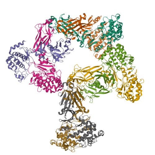
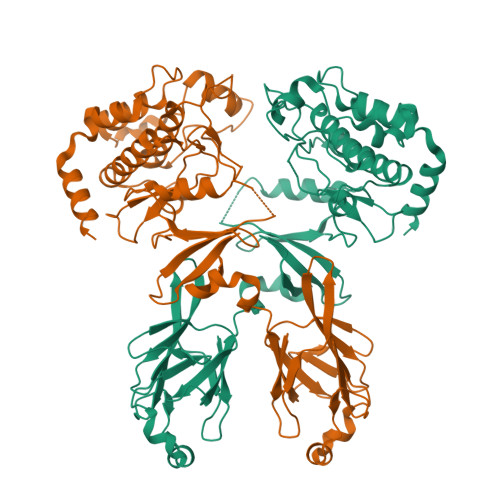
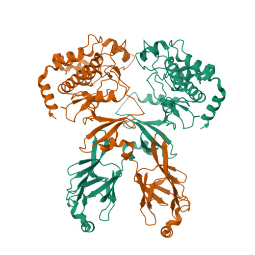
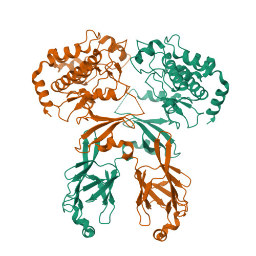
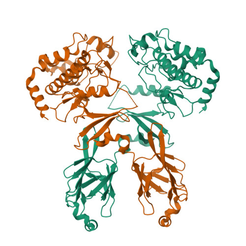
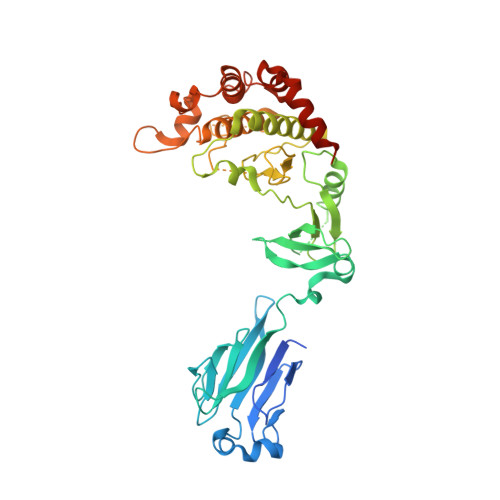
3I6W

Structure and Activation Mechanism of the CHK2 DNA-Damage Checkpoint Kinase


Experimental Data Snapshot

  • Method: X-RAY DIFFRACTION
  • Resolution: 3.25 Å
  • R-Value Free: 0.287 
  • R-Value Work: 0.251 
  • R-Value Observed: 0.252 

Starting Model: experimental
View more details

wwPDB Validation   3D Report Full Report


This is version 1.3 of the entry. See complete history


Literature

Structure and activation mechanism of the CHK2 DNA damage checkpoint kinase.

Cai, Z.Chehab, N.H.Pavletich, N.P.

(2009) Mol Cell 35: 818-829

  • DOI: https://doi.org/10.1016/j.molcel.2009.09.007
  • Primary Citation of Related Structures:  
    3I6U, 3I6W

  • PubMed Abstract: 

    The CHK2 protein kinase is an important transducer of DNA damage checkpoint signals, and its mutation contributes to hereditary and sporadic cancer. CHK2 activation is triggered by the phosphorylation of Thr68 by the DNA damage-activated ATM kinase. This leads to transient CHK2 dimerization, in part through intermolecular phosphoThr68-FHA domain interactions. Dimerization promotes kinase activation through activation-loop autophosphorylation, but the mechanism of this process has not been clear. The dimeric crystal structure of CHK2, described here, in conjunction with biochemical and mutational data reveals that productive CHK2 dimerization additionally involves intermolecular FHA-kinase domain and FHA-FHA interactions. Ile157, mutated in the Li-Fraumeni cancer-predisposition syndrome, plays a central role in the FHA-kinase domain interface, explaining the lack of dimerization and autophosphorylation of this mutant. In the dimer, the kinase active sites face each other in close proximity, indicating that dimerization may also serve to optimally position the kinase active sites for efficient activation loop transphosphorylation.


  • Organizational Affiliation

    Structural Biology Program, Memorial Sloan-Kettering Cancer Center, New York, NY 10021, USA.


Macromolecules
Find similar proteins by:  (by identity cutoff)  |  3D Structure
Entity ID: 1
MoleculeChains Sequence LengthOrganismDetailsImage
Serine/threonine-protein kinase Chk2
A, B, C, D, E
A, B, C, D, E, F, G, H
443Homo sapiensMutation(s): 1 
Gene Names: CHEK2CHK2RAD53
EC: 2.7.11.1
UniProt & NIH Common Fund Data Resources
Find proteins for O96017 (Homo sapiens)
Explore O96017 
Go to UniProtKB:  O96017
PHAROS:  O96017
GTEx:  ENSG00000183765 
Entity Groups  
Sequence Clusters30% Identity50% Identity70% Identity90% Identity95% Identity100% Identity
UniProt GroupO96017
Sequence Annotations
Expand
  • Reference Sequence
Experimental Data & Validation

Experimental Data

  • Method: X-RAY DIFFRACTION
  • Resolution: 3.25 Å
  • R-Value Free: 0.287 
  • R-Value Work: 0.251 
  • R-Value Observed: 0.252 
  • Space Group: P 1
Unit Cell:
Length ( Å )Angle ( ˚ )
a = 76.2α = 84.1
b = 114.7β = 81.2
c = 123γ = 80.7
Software Package:
Software NamePurpose
REFMACrefinement

Structure Validation

View Full Validation Report


Entry History 

Deposition Data

Revision History  (Full details and data files)

  • Version 1.0: 2009-11-10
    Type: Initial release
  • Version 1.1: 2011-07-13
    Changes: Advisory, Version format compliance
  • Version 1.2: 2021-10-13
    Changes: Database references
  • Version 1.3: 2023-09-06
    Changes: Data collection, Refinement description