3QT2 | pdb_00003qt2

Structure of a cytokine ligand-receptor complex


Experimental Data Snapshot

  • Method: X-RAY DIFFRACTION
  • Resolution: 2.55 Å
  • R-Value Free: 
    0.264 (Depositor) 
  • R-Value Work: 
    0.207 (Depositor), 0.210 (DCC) 
  • R-Value Observed: 
    0.210 (Depositor) 

wwPDB Validation   3D Report Full Report


Ligand Structure Quality Assessment 

Created with Raphaël 2.3.0Worse 01 BetterLigand structure goodness of fit to experimental dataBest fitted BGCClick on this verticalbar to view details

This is version 1.3 of the entry. See complete history


Literature

Structure Analysis of the IL-5 Ligand-Receptor Complex Reveals a Wrench-like Architecture for IL-5Ralpha.

Patino, E.Kotzsch, A.Saremba, S.Nickel, J.Schmitz, W.Sebald, W.Mueller, T.D.

(2011) Structure 19: 1864-1875

  • DOI: https://doi.org/10.1016/j.str.2011.08.015
  • Primary Citation of Related Structures:  
    3QT2

  • PubMed Abstract: 

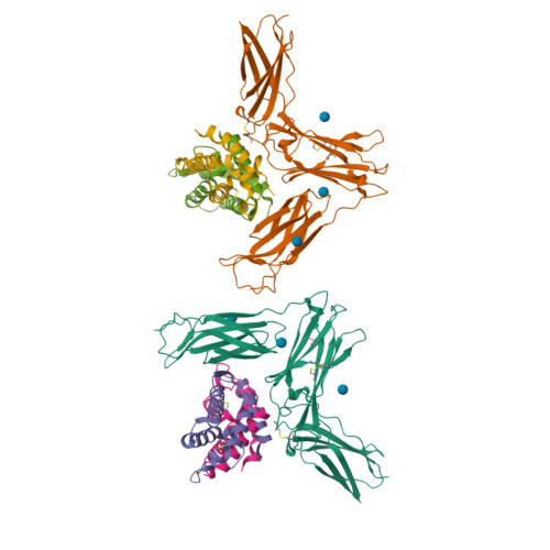
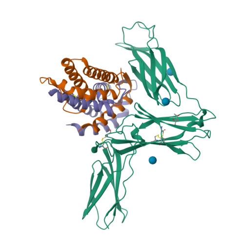
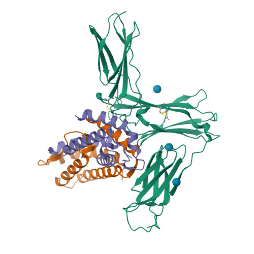
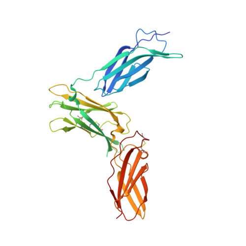
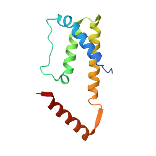
    Interleukin-5 (IL-5) is the key mediator for the function of eosinophil granulocytes, whose deregulation is characteristic of hypereosinophilic diseases and presumably contributes to allergic asthma. IL-5 signaling involves two transmembrane receptors, IL-5Rα and the common β chain, which upon formation of the ternary complex activate the JAK/STAT signaling cascade. To investigate the mechanism underlying ligand-receptor recognition, we determined the structure of IL-5 bound to the extracellular domain of IL-5Rα. IL-5 makes contact with all three fibronectin III-like domains of IL-5Rα, with the receptor architecture resembling a wrench. Mutagenesis data provide evidence that this wrench-like architecture is likely preformed. The structure demonstrates that for steric reasons, homodimeric IL-5 can bind only one receptor molecule, even though two equivalent receptor-binding sites exist. In regard to strong efforts being made to develop IL-5 antagonists for treating asthma and hypereosinophilic diseases, the advances in molecular understanding provided by this structure are of greatest value.


  • Organizational Affiliation

    Lehrstuhl für Molekulare Pflanzenphysiologie und Biophysik, Julius-von-Sachs Institut der Universität Würzburg, Julius-von-Sachs Platz 2, D-97082 Würzburg, Germany.


Macromolecules
Find similar proteins by:  (by identity cutoff)  |  3D Structure
Entity ID: 1
MoleculeChains Sequence LengthOrganismDetailsImage
Interleukin-5 receptor subunit alphaA,
D [auth B]
317Homo sapiensMutation(s): 5 
Gene Names: IL5RIL5RA
UniProt & NIH Common Fund Data Resources
Find proteins for Q01344 (Homo sapiens)
Explore Q01344 
Go to UniProtKB:  Q01344
PHAROS:  Q01344
GTEx:  ENSG00000091181 
Entity Groups  
Sequence Clusters30% Identity50% Identity70% Identity90% Identity95% Identity100% Identity
UniProt GroupQ01344
Sequence Annotations
Expand
  • Reference Sequence
Find similar proteins by:  (by identity cutoff)  |  3D Structure
Entity ID: 2
MoleculeChains Sequence LengthOrganismDetailsImage
Interleukin-5B [auth C],
C [auth D],
E,
F
117Homo sapiensMutation(s): 0 
Gene Names: IL5
UniProt & NIH Common Fund Data Resources
Find proteins for P05113 (Homo sapiens)
Explore P05113 
Go to UniProtKB:  P05113
PHAROS:  P05113
GTEx:  ENSG00000113525 
Entity Groups  
Sequence Clusters30% Identity50% Identity70% Identity90% Identity95% Identity100% Identity
UniProt GroupP05113
Sequence Annotations
Expand
  • Reference Sequence
Small Molecules
Experimental Data & Validation

Experimental Data

  • Method: X-RAY DIFFRACTION
  • Resolution: 2.55 Å
  • R-Value Free:  0.264 (Depositor) 
  • R-Value Work:  0.207 (Depositor), 0.210 (DCC) 
  • R-Value Observed: 0.210 (Depositor) 
Space Group: P 1 21 1
Unit Cell:
Length ( Å )Angle ( ˚ )
a = 85.084α = 90
b = 61.633β = 99.49
c = 142.113γ = 90
Software Package:
Software NamePurpose
SHARPphasing
REFMACrefinement
CrystalCleardata reduction
CrystalCleardata scaling

Structure Validation

View Full Validation Report


Ligand Structure Quality Assessment 

Created with Raphaël 2.3.0Worse 01 BetterLigand structure goodness of fit to experimental dataBest fitted BGCClick on this verticalbar to view details

Entry History 

Deposition Data

Revision History  (Full details and data files)

  • Version 1.0: 2012-02-01
    Type: Initial release
  • Version 1.1: 2017-11-08
    Changes: Refinement description
  • Version 1.2: 2020-07-29
    Type: Remediation
    Reason: Carbohydrate remediation
    Changes: Data collection, Database references, Derived calculations, Structure summary
  • Version 1.3: 2024-10-30
    Changes: Data collection, Database references, Structure summary