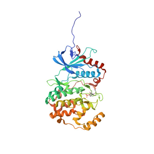
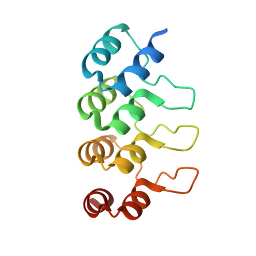
3ZUV

Crystal structure of a designed selected Ankyrin Repeat protein in complex with the phosphorylated MAP kinase ERK2


Experimental Data Snapshot

  • Method: X-RAY DIFFRACTION
  • Resolution: 2.72 Å
  • R-Value Free: 0.230 
  • R-Value Work: 0.176 
  • R-Value Observed: 0.178 

Starting Models: experimental
View more details

wwPDB Validation   3D Report Full Report


This is version 1.3 of the entry. See complete history


Literature

Structural and Functional Analysis of Phosphorylation-Specific Binders of the Kinase Erk from Designed Ankyrin Repeat Protein Libraries.

Kummer, L.Parizek, P.Rube, P.Millgramm, B.Prinz, A.Mittl, P.R.Kaufholz, M.Zimmermann, B.Herberg, F.W.Pluckthun, A.

(2012) Proc Natl Acad Sci U S A 109: E2248

  • DOI: https://doi.org/10.1073/pnas.1205399109
  • Primary Citation of Related Structures:  
    3ZU7, 3ZUV

  • PubMed Abstract: 

    We have selected designed ankyrin repeat proteins (DARPins) from a synthetic library by using ribosome display that selectively bind to the mitogen-activated protein kinase ERK2 (extracellular signal-regulated kinase 2) in either its nonphosphorylated (inactive) or doubly phosphorylated (active) form. They do not bind to other kinases tested. Crystal structures of complexes with two DARPins, each specific for one of the kinase forms, were obtained. The two DARPins bind to essentially the same region of the kinase, but recognize the conformational change within the activation loop and an adjacent area, which is the key structural difference that occurs upon activation. Whereas the rigid phosphorylated activation loop remains in the same form when bound by the DARPin, the more mobile unphosphorylated loop is pushed to a new position. The DARPins can be used to selectively precipitate the cognate form of the kinases from cell lysates. They can also specifically recognize the modification status of the kinase inside the cell. By fusing the kinase with Renilla luciferase and the DARPin to GFP, an energy transfer from luciferase to GFP can be observed in COS-7 cells upon intracellular complex formation. Phosphorylated ERK2 is seen to increase by incubation of the COS-7 cells with FBS and to decrease upon adding the ERK pathway inhibitor PD98509. Furthermore, the anti-ERK2 DARPin is seen to inhibit ERK phosphorylation as it blocks the target inside the cell. This strategy of creating activation-state-specific sensors and kinase-specific inhibitors may add to the repertoire to investigate intracellular signaling in real time.


  • Organizational Affiliation

    Department of Biochemistry, University of Zurich, 8057 Zurich, Switzerland.


Macromolecules
Find similar proteins by:  (by identity cutoff)  |  3D Structure
Entity ID: 1
MoleculeChains Sequence LengthOrganismDetailsImage
MITOGEN-ACTIVATED PROTEIN KINASE 1
A, C
364Rattus norvegicusMutation(s): 0 
EC: 2.7.11.24
UniProt
Find proteins for P63086 (Rattus norvegicus)
Explore P63086 
Go to UniProtKB:  P63086
Entity Groups  
Sequence Clusters30% Identity50% Identity70% Identity90% Identity95% Identity100% Identity
UniProt GroupP63086
Sequence Annotations
Expand
  • Reference Sequence
Find similar proteins by:  (by identity cutoff)  |  3D Structure
Entity ID: 2
MoleculeChains Sequence LengthOrganismDetailsImage
DESIGNED ANKYRIN REPEAT PROTEIN
B, D
136synthetic constructMutation(s): 0 
Entity Groups  
Sequence Clusters30% Identity50% Identity70% Identity90% Identity95% Identity100% Identity
Sequence Annotations
Expand
  • Reference Sequence
Small Molecules
Modified Residues  2 Unique
IDChains TypeFormula2D DiagramParent
PTR
Query on PTR
A, C
L-PEPTIDE LINKINGC9 H12 N O6 PTYR
TPO
Query on TPO
A, C
L-PEPTIDE LINKINGC4 H10 N O6 PTHR
Experimental Data & Validation

Experimental Data

  • Method: X-RAY DIFFRACTION
  • Resolution: 2.72 Å
  • R-Value Free: 0.230 
  • R-Value Work: 0.176 
  • R-Value Observed: 0.178 
  • Space Group: P 21 21 21
Unit Cell:
Length ( Å )Angle ( ˚ )
a = 113.67α = 90
b = 150.449β = 90
c = 104.97γ = 90
Software Package:
Software NamePurpose
REFMACrefinement
XDSdata reduction
XSCALEdata scaling
PHASERphasing

Structure Validation

View Full Validation Report


Entry History 

Deposition Data

Revision History  (Full details and data files)

  • Version 1.0: 2012-06-27
    Type: Initial release
  • Version 1.1: 2012-09-05
    Changes: Database references
  • Version 1.2: 2023-12-20
    Changes: Data collection, Database references, Derived calculations, Other, Refinement description
  • Version 1.3: 2024-11-13
    Changes: Structure summary