4NFF | pdb_00004nff

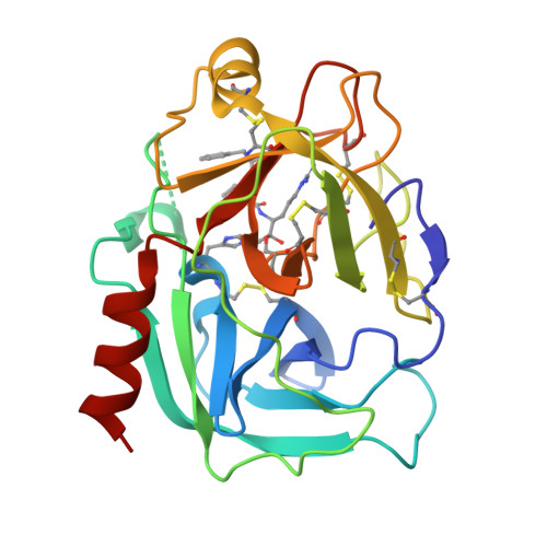
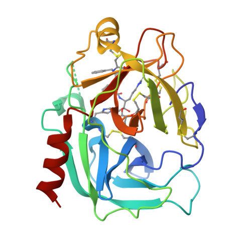
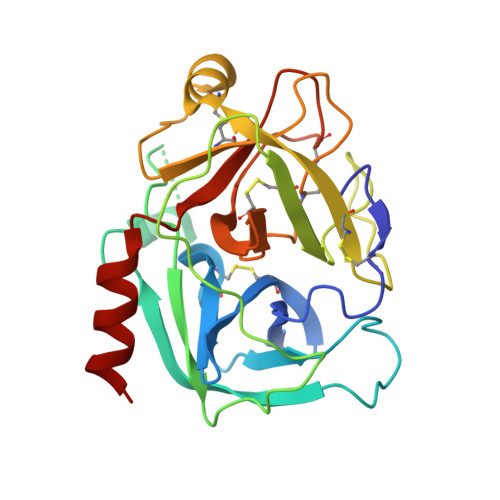
Human kallikrein-related peptidase 2 in complex with PPACK


Experimental Data Snapshot

  • Method: X-RAY DIFFRACTION
  • Resolution: 1.90 Å
  • R-Value Free: 
    0.232 (Depositor), 0.233 (DCC) 
  • R-Value Work: 
    0.197 (Depositor), 0.197 (DCC) 
  • R-Value Observed: 
    0.199 (Depositor) 

wwPDB Validation   3D Report Full Report


Ligand Structure Quality Assessment 

Created with Raphaël 2.3.0Worse 01 BetterLigand structure goodness of fit to experimental dataBest fitted 0G6Click on this verticalbar to view details

This is version 1.3 of the entry. See complete history


Literature

Structure-function analyses of human kallikrein-related peptidase 2 establish the 99-loop as master regulator of activity

Skala, W.Utzschneider, D.T.Magdolen, V.Debela, M.Guo, S.Craik, C.S.Brandstetter, H.Goettig, P.

(2014) J Biological Chem 289: 34267-34283

  • DOI: https://doi.org/10.1074/jbc.M114.598201
  • Primary Citation of Related Structures:  
    4NFE, 4NFF

  • PubMed Abstract: 

    Human kallikrein-related peptidase 2 (KLK2) is a tryptic serine protease predominantly expressed in prostatic tissue and secreted into prostatic fluid, a major component of seminal fluid. Most likely it activates and complements chymotryptic KLK3 (prostate-specific antigen) in cleaving seminal clotting proteins, resulting in sperm liquefaction. KLK2 belongs to the "classical" KLKs 1-3, which share an extended 99- or kallikrein loop near their non-primed substrate binding site. Here, we report the 1.9 Å crystal structures of two KLK2-small molecule inhibitor complexes. In both structures discontinuous electron density for the 99-loop indicates that this loop is largely disordered. We provide evidence that the 99-loop is responsible for two biochemical peculiarities of KLK2, i.e. reversible inhibition by micromolar Zn(2+) concentrations and permanent inactivation by autocatalytic cleavage. Indeed, several 99-loop mutants of KLK2 displayed an altered susceptibility to Zn(2+), which located the Zn(2+) binding site at the 99-loop/active site interface. In addition, we identified an autolysis site between residues 95e and 95f in the 99-loop, whose elimination prevented the mature enzyme from limited autolysis and irreversible inactivation. An exhaustive comparison of KLK2 with related structures revealed that in the KLK family the 99-, 148-, and 220-loop exist in open and closed conformations, allowing or preventing substrate access, which extends the concept of conformational selection in trypsin-related proteases. Taken together, our novel biochemical and structural data on KLK2 identify its 99-loop as a key player in activity regulation.


  • Organizational Affiliation

    From the Division of Structural Biology, Department of Molecular Biology, University of Salzburg, A-5020 Salzburg, Austria.


Macromolecules
Find similar proteins by:  (by identity cutoff)  |  3D Structure
Entity ID: 1
MoleculeChains Sequence LengthOrganismDetailsImage
Kallikrein-2237Homo sapiensMutation(s): 0 
Gene Names: KLK2
EC: 3.4.21.35
UniProt & NIH Common Fund Data Resources
Find proteins for P20151 (Homo sapiens)
Explore P20151 
Go to UniProtKB:  P20151
PHAROS:  P20151
GTEx:  ENSG00000167751 
Entity Groups  
Sequence Clusters30% Identity50% Identity70% Identity90% Identity95% Identity100% Identity
UniProt GroupP20151
Sequence Annotations
Expand
  • Reference Sequence
Small Molecules
Ligands 1 Unique
IDChains Name / Formula / InChI Key2D Diagram3D Interactions
0G6
Query on 0G6

Download Ideal Coordinates CCD File 
B [auth A]D-phenylalanyl-N-[(2S,3S)-6-{[amino(iminio)methyl]amino}-1-chloro-2-hydroxyhexan-3-yl]-L-prolinamide
C21 H34 Cl N6 O3
DVFLYEYCMMLBTQ-VSZNYVQBSA-O
Biologically Interesting Molecules (External Reference) 1 Unique
Experimental Data & Validation

Experimental Data

  • Method: X-RAY DIFFRACTION
  • Resolution: 1.90 Å
  • R-Value Free:  0.232 (Depositor), 0.233 (DCC) 
  • R-Value Work:  0.197 (Depositor), 0.197 (DCC) 
  • R-Value Observed: 0.199 (Depositor) 
Space Group: P 21 21 21
Unit Cell:
Length ( Å )Angle ( ˚ )
a = 60.1α = 90
b = 60.74β = 90
c = 66.8γ = 90
Software Package:
Software NamePurpose
MOSFLMdata reduction
SCALAdata scaling
PHASERphasing
REFMACrefinement
PDB_EXTRACTdata extraction
MAR345dtbdata collection

Structure Validation

View Full Validation Report


Ligand Structure Quality Assessment 

Created with Raphaël 2.3.0Worse 01 BetterLigand structure goodness of fit to experimental dataBest fitted 0G6Click on this verticalbar to view details

Entry History 

Deposition Data

Revision History  (Full details and data files)

  • Version 1.0: 2014-10-29
    Type: Initial release
  • Version 1.1: 2015-04-08
    Changes: Database references
  • Version 1.2: 2017-11-15
    Changes: Refinement description
  • Version 1.3: 2024-10-30
    Changes: Data collection, Database references, Derived calculations, Structure summary