4NQA

Crystal structure of liganded hRXR-alpha/hLXR-beta heterodimer on DNA


Experimental Data Snapshot

  • Method: X-RAY DIFFRACTION
  • Resolution: 3.10 Å
  • R-Value Free: 0.218 
  • R-Value Work: 0.180 
  • R-Value Observed: 0.186 

wwPDB Validation   3D Report Full Report


Ligand Structure Quality Assessment 


This is version 1.3 of the entry. See complete history


Literature

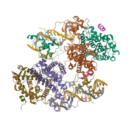
Structure of the retinoid X receptor alpha-liver X receptor beta (RXR alpha-LXR beta ) heterodimer on DNA.

Lou, X.Toresson, G.Benod, C.Suh, J.H.Philips, K.J.Webb, P.Gustafsson, J.A.

(2014) Nat Struct Mol Biol 21: 277-281

  • DOI: https://doi.org/10.1038/nsmb.2778
  • Primary Citation of Related Structures:  
    4NQA

  • PubMed Abstract: 

    Nuclear receptors (NRs) are conditional transcription factors with common multidomain organization that bind diverse DNA elements. How DNA sequences influence NR conformation is poorly understood. Here we report the crystal structure of the human retinoid X receptor α-liver X receptor β (RXRα-LXRβ) heterodimer on its cognate element, an AGGTCA direct repeat spaced by 4 nt. The complex has an extended X-shaped arrangement, with DNA- and ligand-binding domains crossed, in contrast to the parallel domain arrangement of other NRs that bind an AGGTCA direct repeat spaced by 1 nt. The LXRβ core binds DNA via canonical contacts and auxiliary DNA contacts that enhance affinity for the response element. Comparisons of RXRα-LXRβs in the crystal asymmetric unit and with previous NR structures reveal flexibility in NR organization and suggest a role for RXRα in adaptation of heterodimeric complexes to DNA.


  • Organizational Affiliation

    1] Center for Nuclear Receptors and Cell Signaling, University of Houston, Houston, Texas, USA. [2] Genomic Medicine Program, Houston Methodist Research Institute, Houston, Texas, USA.


Macromolecules

Find similar proteins by:  (by identity cutoff)  |  3D Structure
Entity ID: 1
MoleculeChains Sequence LengthOrganismDetailsImage
Retinoic acid receptor RXR-alphaA,
G [auth H]
365Homo sapiensMutation(s): 0 
Gene Names: RXRANR2B1
UniProt & NIH Common Fund Data Resources
Find proteins for P19793 (Homo sapiens)
Explore P19793 
Go to UniProtKB:  P19793
PHAROS:  P19793
GTEx:  ENSG00000186350 
Entity Groups  
Sequence Clusters30% Identity50% Identity70% Identity90% Identity95% Identity100% Identity
UniProt GroupP19793
Sequence Annotations
Expand
  • Reference Sequence
Find similar proteins by:  (by identity cutoff)  |  3D Structure
Entity ID: 2
MoleculeChains Sequence LengthOrganismDetailsImage
Liver X nuclear receptor betaB,
H [auth I]
391Homo sapiensMutation(s): 0 
Gene Names: NR1H2hCG_22944
UniProt & NIH Common Fund Data Resources
Find proteins for P55055 (Homo sapiens)
Explore P55055 
Go to UniProtKB:  P55055
PHAROS:  P55055
GTEx:  ENSG00000131408 
Entity Groups  
Sequence Clusters30% Identity50% Identity70% Identity90% Identity95% Identity100% Identity
UniProt GroupP55055
Sequence Annotations
Expand
  • Reference Sequence

Find similar proteins by:  Sequence   |   3D Structure  

Entity ID: 3
MoleculeChains Sequence LengthOrganismDetailsImage
Nuclear receptor coactivator 2C,
D,
I [auth J],
J [auth K]
13Homo sapiensMutation(s): 0 
Gene Names: NCOA2BHLHE75TIF2
UniProt & NIH Common Fund Data Resources
Find proteins for Q15596 (Homo sapiens)
Explore Q15596 
Go to UniProtKB:  Q15596
PHAROS:  Q15596
GTEx:  ENSG00000140396 
Entity Groups  
Sequence Clusters30% Identity50% Identity70% Identity90% Identity95% Identity100% Identity
UniProt GroupQ15596
Sequence Annotations
Expand
  • Reference Sequence

Find similar nucleic acids by:  Sequence   |   3D Structure  

Entity ID: 4
MoleculeChains LengthOrganismImage
5'-D(*TP*AP*AP*GP*GP*TP*CP*AP*CP*TP*TP*CP*AP*GP*GP*TP*CP*A)-3'E,
K [auth L]
18synthetic construct
Sequence Annotations
Expand
  • Reference Sequence

Find similar nucleic acids by:  Sequence   |   3D Structure  

Entity ID: 5
MoleculeChains LengthOrganismImage
5'-D(*TP*AP*TP*GP*AP*CP*CP*TP*GP*AP*AP*GP*TP*GP*AP*CP*CP*T)-3'F,
L [auth M]
18synthetic construct
Sequence Annotations
Expand
  • Reference Sequence
Small Molecules
Ligands 3 Unique
IDChains Name / Formula / InChI Key2D Diagram3D Interactions
965
Query on 965

Download Ideal Coordinates CCD File 
P [auth B],
V [auth I]
[3-(3-{[2-chloro-3-(trifluoromethyl)benzyl](2,2-diphenylethyl)amino}propoxy)phenyl]acetic acid
C33 H31 Cl F3 N O3
NAXSRXHZFIBFMI-UHFFFAOYSA-N
9CR
Query on 9CR

Download Ideal Coordinates CCD File 
M [auth A],
S [auth H]
(9cis)-retinoic acid
C20 H28 O2
SHGAZHPCJJPHSC-ZVCIMWCZSA-N
ZN
Query on ZN

Download Ideal Coordinates CCD File 
N [auth A]
O [auth A]
Q [auth B]
R [auth B]
T [auth H]
N [auth A],
O [auth A],
Q [auth B],
R [auth B],
T [auth H],
U [auth H],
W [auth I],
X [auth I]
ZINC ION
Zn
PTFCDOFLOPIGGS-UHFFFAOYSA-N
Binding Affinity Annotations 
IDSourceBinding Affinity
965 BindingDB:  4NQA Ki: 2.2 (nM) from 1 assay(s)
IC50: min: 7.9, max: 40 (nM) from 5 assay(s)
EC50: min: 5, max: 501 (nM) from 15 assay(s)
9CR BindingDB:  4NQA Ki: min: 8, max: 583 (nM) from 5 assay(s)
Kd: min: 1.5, max: 1810 (nM) from 11 assay(s)
IC50: min: 29, max: 82 (nM) from 3 assay(s)
EC50: min: 1.5, max: 1730 (nM) from 14 assay(s)
Experimental Data & Validation

Experimental Data

  • Method: X-RAY DIFFRACTION
  • Resolution: 3.10 Å
  • R-Value Free: 0.218 
  • R-Value Work: 0.180 
  • R-Value Observed: 0.186 
  • Space Group: P 43
Unit Cell:
Length ( Å )Angle ( ˚ )
a = 85.783α = 90
b = 85.783β = 90
c = 238.577γ = 90
Software Package:
Software NamePurpose
HKL-2000data collection
PHASESphasing
PHENIXrefinement
HKL-2000data reduction
HKL-2000data scaling

Structure Validation

View Full Validation Report


Ligand Structure Quality Assessment 


Entry History 

Deposition Data

Revision History  (Full details and data files)

  • Version 1.0: 2014-02-26
    Type: Initial release
  • Version 1.1: 2014-03-19
    Changes: Database references
  • Version 1.2: 2016-11-09
    Changes: Non-polymer description, Source and taxonomy
  • Version 1.3: 2024-02-28
    Changes: Data collection, Database references, Derived calculations