Structural Basis for Inhibition of Mycobacterial and Human Adenosine Kinase by 7-Substituted 7-(Het)aryl-7-deazaadenine Ribonucleosides
Snasel, J., Naus, P., Dostal, J., Hnizda, A., Fanfrlik, J., Brynda, J., Bourderioux, A., Dusek, M., Dvorakova, H., Stolarikova, J., Zabranska, H., Pohl, R., Konecny, P., Dzubak, P., Votruba, I., Hajduch, M., Rezacova, P., Veverka, V., Hocek, M., Pichova, I.(2014) J Med Chem 57: 8268-8279
- PubMed: 25259627
- DOI: https://doi.org/10.1021/jm500497v
- Primary Citation of Related Structures:
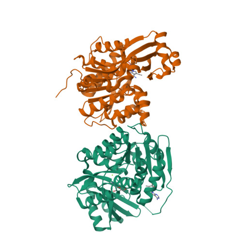
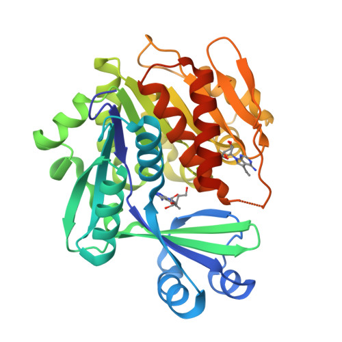
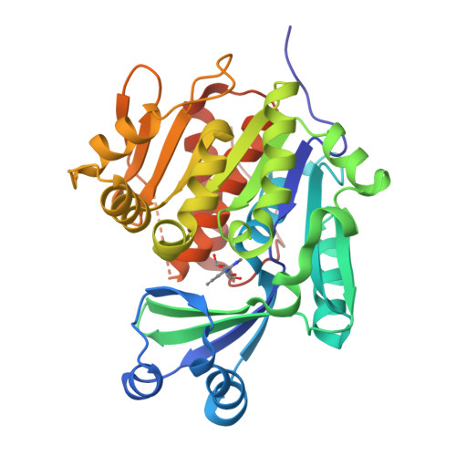
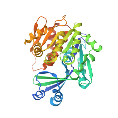
4O1G, 4O1L, 4PVV - PubMed Abstract:
Adenosine kinase (ADK) from Mycobacterium tuberculosis (Mtb) was selected as a target for design of antimycobacterial nucleosides. Screening of 7-(het)aryl-7-deazaadenine ribonucleosides with Mtb and human (h) ADKs and testing with wild-type and drug-resistant Mtb strains identified specific inhibitors of Mtb ADK with micromolar antimycobacterial activity and low cytotoxicity. X-ray structures of complexes of Mtb and hADKs with 7-ethynyl-7-deazaadenosine showed differences in inhibitor interactions in the adenosine binding sites. 1D (1)H STD NMR experiments revealed that these inhibitors are readily accommodated into the ATP and adenosine binding sites of Mtb ADK, whereas they bind preferentially into the adenosine site of hADK. Occupation of the Mtb ADK ATP site with inhibitors and formation of catalytically less competent semiopen conformation of MtbADK after inhibitor binding in the adenosine site explain the lack of phosphorylation of 7-substituted-7-deazaadenosines. Semiempirical quantum mechanical analysis confirmed different affinity of nucleosides for the Mtb ADK adenosine and ATP sites.
Organizational Affiliation:
Institute of Organic Chemistry and Biochemistry, Academy of Sciences of the Czech Republic , Flemingovo nam. 2, 16610 Prague 6, Czech Republic.