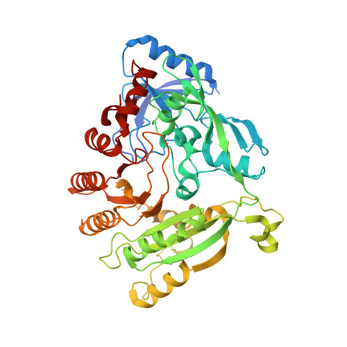
2-Carboxyquinoxalines Kill Mycobacterium tuberculosis through Noncovalent Inhibition of DprE1.
Neres, J., Hartkoorn, R.C., Chiarelli, L.R., Gadupudi, R., Pasca, M.R., Mori, G., Venturelli, A., Savina, S., Makarov, V., Kolly, G.S., Molteni, E., Binda, C., Dhar, N., Ferrari, S., Brodin, P., Delorme, V., Landry, V., de Jesus Lopes Ribeiro, A.L., Farina, D., Saxena, P., Pojer, F., Carta, A., Luciani, R., Porta, A., Zanoni, G., De Rossi, E., Costi, M.P., Riccardi, G., Cole, S.T.(2015) ACS Chem Biol 10: 705-714
- PubMed: 25427196
- DOI: https://doi.org/10.1021/cb5007163
- Primary Citation of Related Structures:
4CVY, 4P8C, 4P8K, 4P8L, 4P8M, 4P8N, 4P8P, 4P8T, 4P8Y - PubMed Abstract:
Phenotypic screening of a quinoxaline library against replicating Mycobacterium tuberculosis led to the identification of lead compound Ty38c (3-((4-methoxybenzyl)amino)-6-(trifluoromethyl)quinoxaline-2-carboxylic acid). With an MIC99 and MBC of 3.1 μM, Ty38c is bactericidal and active against intracellular bacteria. To investigate its mechanism of action, we isolated mutants resistant to Ty38c and sequenced their genomes. Mutations were found in rv3405c, coding for the transcriptional repressor of the divergently expressed rv3406 gene. Biochemical studies clearly showed that Rv3406 decarboxylates Ty38c into its inactive keto metabolite. The actual target was then identified by isolating Ty38c-resistant mutants of an M. tuberculosis strain lacking rv3406. Here, mutations were found in dprE1, encoding the decaprenylphosphoryl-d-ribose oxidase DprE1, essential for biogenesis of the mycobacterial cell wall. Genetics, biochemical validation, and X-ray crystallography revealed Ty38c to be a noncovalent, noncompetitive DprE1 inhibitor. Structure-activity relationship studies generated a family of DprE1 inhibitors with a range of IC50's and bactericidal activity. Co-crystal structures of DprE1 in complex with eight different quinoxaline analogs provided a high-resolution interaction map of the active site of this extremely vulnerable target in M. tuberculosis.
Organizational Affiliation:
†More Medicines for Tuberculosis (MM4TB) Consortium (www.mm4tb.org).


