4UUU | pdb_00004uuu

1.7 A resolution structure of human cystathionine beta-synthase regulatory domain (del 516-525) in complex with SAM


Experimental Data Snapshot

  • Method: X-RAY DIFFRACTION
  • Resolution: 1.71 Å
  • R-Value Free: 
    0.199 (Depositor), 0.210 (DCC) 
  • R-Value Work: 
    0.159 (Depositor), 0.170 (DCC) 
  • R-Value Observed: 
    0.161 (Depositor) 

wwPDB Validation   3D Report Full Report


Ligand Structure Quality Assessment 

Created with Raphaël 2.3.0Worse 01 BetterLigand structure goodness of fit to experimental dataBest fitted SAMClick on this verticalbar to view details

This is version 1.5 of the entry. See complete history


Literature

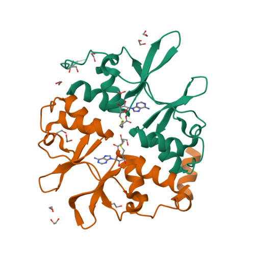
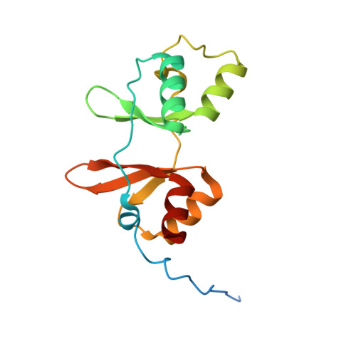
Inter-Domain Communication of Human Cystathionine Beta Synthase: Structural Basis of S-Adenosyl-L-Methionine Activation.

Mccorvie, T.J.Kopec, J.Hyung, S.Fitzpatrick, F.Feng, X.Termine, D.Strain-Damerell, C.Vollmar, M.Fleming, J.Janz, J.M.Bulawa, C.Yue, W.W.

(2014) J Biological Chem 289: 36018

  • DOI: https://doi.org/10.1074/jbc.M114.610782
  • Primary Citation of Related Structures:  
    4COO, 4UUU

  • PubMed Abstract: 

    Cystathionine β-synthase (CBS) is a key enzyme in sulfur metabolism, and its inherited deficiency causes homocystinuria. Mammalian CBS is modulated by the binding of S-adenosyl-l-methionine (AdoMet) to its regulatory domain, which activates its catalytic domain. To investigate the underlying mechanism, we performed x-ray crystallography, mutagenesis, and mass spectrometry (MS) on human CBS. The 1.7 Å structure of a AdoMet-bound CBS regulatory domain shows one AdoMet molecule per monomer, at the interface between two constituent modules (CBS-1, CBS-2). AdoMet binding is accompanied by a reorientation between the two modules, relative to the AdoMet-free basal state, to form interactions with AdoMet via residues verified by mutagenesis to be important for AdoMet binding (Phe(443), Asp(444), Gln(445), and Asp(538)) and for AdoMet-driven inter-domain communication (Phe(443), Asp(538)). The observed structural change is further supported by ion mobility MS, showing that as-purified CBS exists in two conformational populations, which converged to one in the presence of AdoMet. We therefore propose that AdoMet-induced conformational change alters the interface and arrangement between the catalytic and regulatory domains within the CBS oligomer, thereby increasing the accessibility of the enzyme active site for catalysis.


  • Organizational Affiliation

    From the Structural Genomics Consortium, Nuffield Department of Clinical Medicine, University of Oxford, Oxford OX3 7DQ, United Kingdom.


Macromolecules
Find similar proteins by:  (by identity cutoff)  |  3D Structure
Entity ID: 1
MoleculeChains Sequence LengthOrganismDetailsImage
CYSTATHIONINE BETA-SYNTHASE
A, B
155Homo sapiensMutation(s): 0 
EC: 4.2.1.22
UniProt & NIH Common Fund Data Resources
Find proteins for P35520 (Homo sapiens)
Explore P35520 
Go to UniProtKB:  P35520
PHAROS:  P35520
GTEx:  ENSG00000160200 
Entity Groups  
Sequence Clusters30% Identity50% Identity70% Identity90% Identity95% Identity100% Identity
UniProt GroupP35520
Sequence Annotations
Expand
  • Reference Sequence
Small Molecules
Ligands 2 Unique
IDChains Name / Formula / InChI Key2D Diagram3D Interactions
SAM
Query on SAM

Download Ideal Coordinates CCD File 
C [auth A],
K [auth B]
S-ADENOSYLMETHIONINE
C15 H22 N6 O5 S
MEFKEPWMEQBLKI-FCKMPRQPSA-N
EDO
Query on EDO

Download Ideal Coordinates CCD File 
D [auth A]
E [auth A]
F [auth A]
G [auth A]
H [auth A]
1,2-ETHANEDIOL
C2 H6 O2
LYCAIKOWRPUZTN-UHFFFAOYSA-N
Experimental Data & Validation

Experimental Data

  • Method: X-RAY DIFFRACTION
  • Resolution: 1.71 Å
  • R-Value Free:  0.199 (Depositor), 0.210 (DCC) 
  • R-Value Work:  0.159 (Depositor), 0.170 (DCC) 
  • R-Value Observed: 0.161 (Depositor) 
Space Group: P 21 21 21
Unit Cell:
Length ( Å )Angle ( ˚ )
a = 39.85α = 90
b = 78.65β = 90
c = 90.8γ = 90
Software Package:
Software NamePurpose
REFMACrefinement
XDSdata reduction
Aimlessdata scaling
SHELXphasing

Structure Validation

View Full Validation Report


Ligand Structure Quality Assessment 

Created with Raphaël 2.3.0Worse 01 BetterLigand structure goodness of fit to experimental dataBest fitted SAMClick on this verticalbar to view details

Entry History 

Revision History  (Full details and data files)

  • Version 1.0: 2014-08-13
    Type: Initial release
  • Version 1.1: 2014-08-20
    Changes: Structure summary
  • Version 1.2: 2014-09-17
    Changes: Structure summary
  • Version 1.3: 2014-11-05
    Changes: Database references
  • Version 1.4: 2015-01-14
    Changes: Database references
  • Version 1.5: 2024-05-08
    Changes: Data collection, Database references, Derived calculations, Other