4WQO

Structure of VHL-EloB-EloC-Cul2


Experimental Data Snapshot

  • Method: X-RAY DIFFRACTION
  • Resolution: 3.20 Å
  • R-Value Free: 0.249 
  • R-Value Work: 0.221 
  • R-Value Observed: 0.222 

wwPDB Validation   3D Report Full Report


This is version 1.3 of the entry. See complete history


Literature

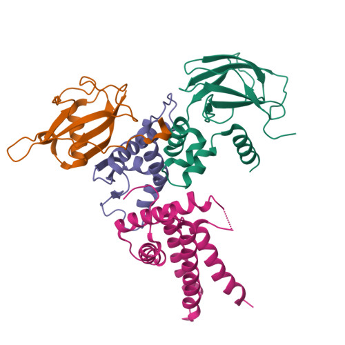
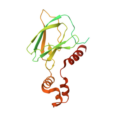
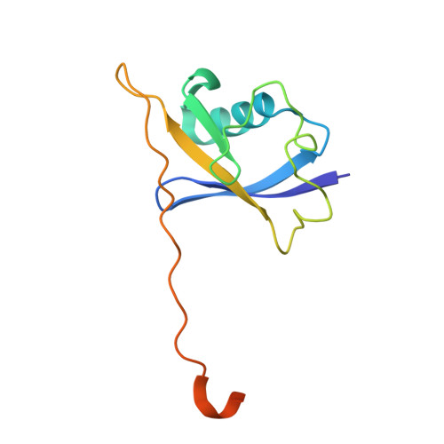
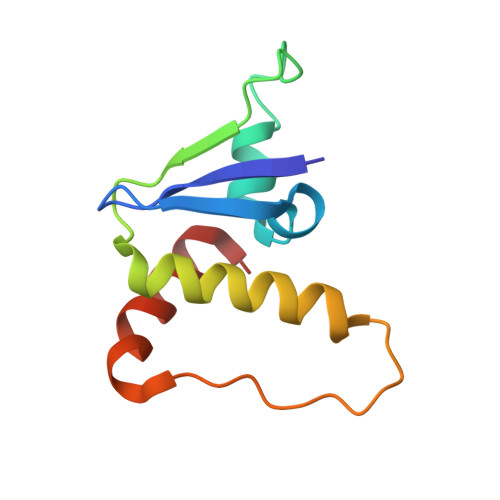
Insights into Cullin-RING E3 Ubiquitin Ligase Recruitment: Structure of the VHL-EloBC-Cul2 Complex.

Nguyen, H.C.Yang, H.Fribourgh, J.L.Wolfe, L.S.Xiong, Y.

(2015) Structure 23: 441-449

  • DOI: https://doi.org/10.1016/j.str.2014.12.014
  • Primary Citation of Related Structures:  
    4WQO

  • PubMed Abstract: 

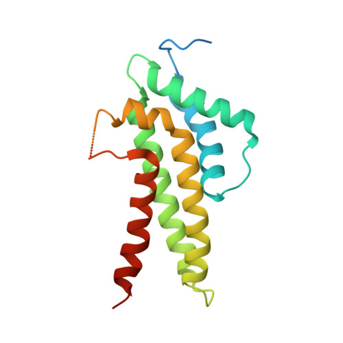
    The von Hippel-Lindau tumor suppressor protein (VHL) recruits a Cullin 2 (Cul2) E3 ubiquitin ligase to downregulate HIF-1α, an essential transcription factor for the hypoxia response. Mutations in VHL lead to VHL disease and renal cell carcinomas. Inhibition of this pathway to upregulate erythropoietin production is a promising new therapy to treat ischemia and chronic anemia. Here, we report the crystal structure of VHL bound to a Cul2 N-terminal domain, Elongin B, and Elongin C (EloC). Cul2 interacts with both the VHL BC box and cullin box and a novel EloC site. Comparison with other cullin E3 ligase structures shows that there is a conserved, yet flexible, cullin recognition module and that cullin selectivity is influenced by distinct electrostatic interactions. Our structure provides a structural basis for the study of the pathogenesis of VHL disease and rationale for the design of novel compounds that may modulate cullin-substrate receptor interactions.


  • Organizational Affiliation

    Department of Molecular Biophysics and Biochemistry, Yale University, New Haven, CT 06511, USA.


Macromolecules
Find similar proteins by:  (by identity cutoff)  |  3D Structure
Entity ID: 1
MoleculeChains Sequence LengthOrganismDetailsImage
Von Hippel-Lindau disease tumor suppressor233Homo sapiensMutation(s): 0 
Gene Names: VHL
UniProt & NIH Common Fund Data Resources
Find proteins for P40337 (Homo sapiens)
Explore P40337 
Go to UniProtKB:  P40337
PHAROS:  P40337
GTEx:  ENSG00000134086 
Entity Groups  
Sequence Clusters30% Identity50% Identity70% Identity90% Identity95% Identity100% Identity
UniProt GroupP40337
Sequence Annotations
Expand
  • Reference Sequence
Find similar proteins by:  (by identity cutoff)  |  3D Structure
Entity ID: 2
MoleculeChains Sequence LengthOrganismDetailsImage
Transcription elongation factor B polypeptide 2118Homo sapiensMutation(s): 0 
Gene Names: TCEB2
UniProt & NIH Common Fund Data Resources
Find proteins for Q15370 (Homo sapiens)
Explore Q15370 
Go to UniProtKB:  Q15370
PHAROS:  Q15370
GTEx:  ENSG00000103363 
Entity Groups  
Sequence Clusters30% Identity50% Identity70% Identity90% Identity95% Identity100% Identity
UniProt GroupQ15370
Sequence Annotations
Expand
  • Reference Sequence
Find similar proteins by:  (by identity cutoff)  |  3D Structure
Entity ID: 3
MoleculeChains Sequence LengthOrganismDetailsImage
Transcription elongation factor B polypeptide 196Homo sapiensMutation(s): 0 
Gene Names: TCEB1
UniProt & NIH Common Fund Data Resources
Find proteins for Q15369 (Homo sapiens)
Explore Q15369 
Go to UniProtKB:  Q15369
PHAROS:  Q15369
GTEx:  ENSG00000154582 
Entity Groups  
Sequence Clusters30% Identity50% Identity70% Identity90% Identity95% Identity100% Identity
UniProt GroupQ15369
Sequence Annotations
Expand
  • Reference Sequence
Find similar proteins by:  (by identity cutoff)  |  3D Structure
Entity ID: 4
MoleculeChains Sequence LengthOrganismDetailsImage
Cullin-2186Homo sapiensMutation(s): 0 
Gene Names: CUL2
UniProt & NIH Common Fund Data Resources
Find proteins for Q13617 (Homo sapiens)
Explore Q13617 
Go to UniProtKB:  Q13617
PHAROS:  Q13617
GTEx:  ENSG00000108094 
Entity Groups  
Sequence Clusters30% Identity50% Identity70% Identity90% Identity95% Identity100% Identity
UniProt GroupQ13617
Sequence Annotations
Expand
  • Reference Sequence
Experimental Data & Validation

Experimental Data

  • Method: X-RAY DIFFRACTION
  • Resolution: 3.20 Å
  • R-Value Free: 0.249 
  • R-Value Work: 0.221 
  • R-Value Observed: 0.222 
  • Space Group: P 32 2 1
Unit Cell:
Length ( Å )Angle ( ˚ )
a = 108.278α = 90
b = 108.278β = 90
c = 213.768γ = 120
Software Package:
Software NamePurpose
PHENIXrefinement
HKL-2000data reduction
SCALEPACKdata scaling
PDB_EXTRACTdata extraction

Structure Validation

View Full Validation Report


Entry History 

Deposition Data

Revision History  (Full details and data files)

  • Version 1.0: 2015-03-04
    Type: Initial release
  • Version 1.1: 2015-03-11
    Changes: Database references
  • Version 1.2: 2017-11-22
    Changes: Database references, Derived calculations, Refinement description, Source and taxonomy
  • Version 1.3: 2023-09-27
    Changes: Data collection, Database references, Refinement description