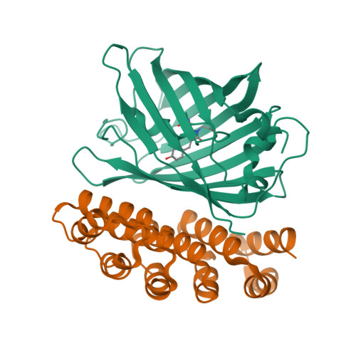
4XVP

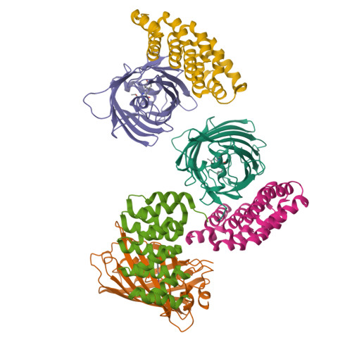
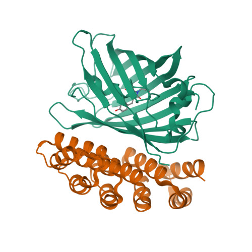
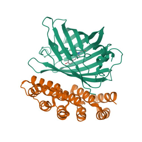
X-ray structure of bGFP-C / EGFP complex


Experimental Data Snapshot

  • Method: X-RAY DIFFRACTION
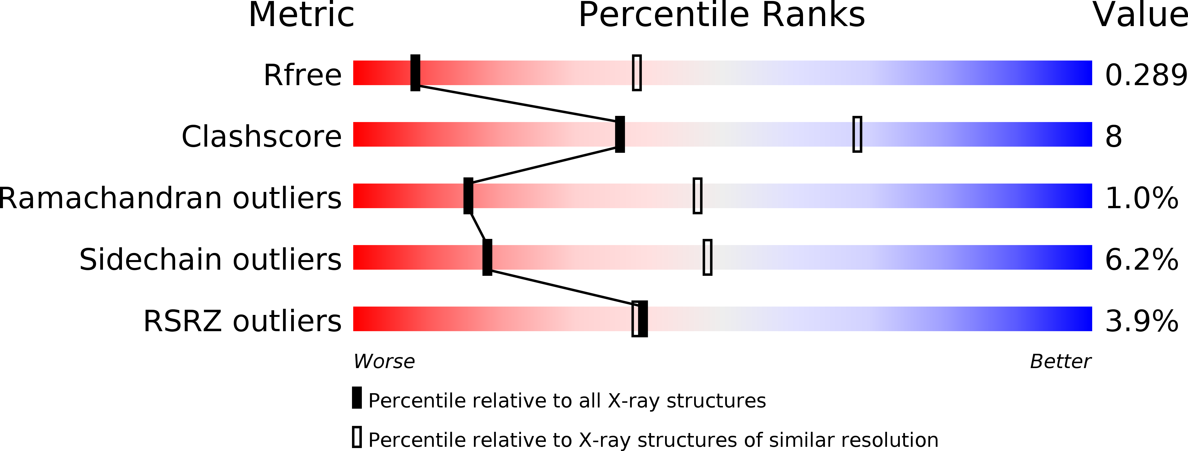
  • Resolution: 3.40 Å
  • R-Value Free: 0.292 
  • R-Value Work: 0.209 
  • R-Value Observed: 0.213 

Starting Models: experimental
View more details

wwPDB Validation   3D Report Full Report


This is version 1.3 of the entry. See complete history


Literature

Specific GFP-binding artificial proteins ( alpha Rep): a new tool for in vitro to live cell applications.

Chevrel, A.Urvoas, A.de la Sierra-Gallay, I.L.Aumont-Nicaise, M.Moutel, S.Desmadril, M.Perez, F.Gautreau, A.van Tilbeurgh, H.Minard, P.Valerio-Lepiniec, M.

(2015) Biosci Rep 35

  • DOI: https://doi.org/10.1042/BSR20150080
  • Primary Citation of Related Structures:  
    4XL5, 4XVP

  • PubMed Abstract: 

    A family of artificial proteins, named αRep, based on a natural family of helical repeat was previously designed. αRep members are efficiently expressed, folded and extremely stable proteins. A large αRep library was constructed creating proteins with a randomized interaction surface. In the present study, we show that the αRep library is an efficient source of tailor-made specific proteins with direct applications in biochemistry and cell biology. From this library, we selected by phage display αRep binders with nanomolar dissociation constants against the GFP. The structures of two independent αRep binders in complex with the GFP target were solved by X-ray crystallography revealing two totally different binding modes. The affinity of the selected αReps for GFP proved sufficient for practically useful applications such as pull-down experiments. αReps are disulfide free proteins and are efficiently and functionally expressed in eukaryotic cells: GFP-specific αReps are clearly sequestrated by their cognate target protein addressed to various cell compartments. These results suggest that αRep proteins with tailor-made specificity can be selected and used in living cells to track, modulate or interfere with intracellular processes.


  • Organizational Affiliation

    Institute for Integrative Biology of the Cell (I2BC), UMR 9198, CEA, CNRS, Université Paris-Sud, Bât 430, 91405 Orsay Cedex, France.


Macromolecules
Find similar proteins by:  (by identity cutoff)  |  3D Structure
Entity ID: 1
MoleculeChains Sequence LengthOrganismDetailsImage
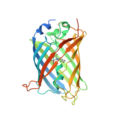
Green fluorescent protein
A, B, C
272Aequorea victoriaMutation(s): 3 
Gene Names: GFP
UniProt
Find proteins for P42212 (Aequorea victoria)
Explore P42212 
Go to UniProtKB:  P42212
Entity Groups  
Sequence Clusters30% Identity50% Identity70% Identity90% Identity95% Identity100% Identity
UniProt GroupP42212
Sequence Annotations
Expand
  • Reference Sequence
Find similar proteins by:  (by identity cutoff)  |  3D Structure
Entity ID: 2
MoleculeChains Sequence LengthOrganismDetailsImage
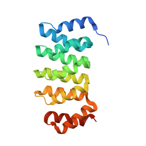
BGFP-C
D, E, F
170synthetic constructMutation(s): 0 
Entity Groups  
Sequence Clusters30% Identity50% Identity70% Identity90% Identity95% Identity100% Identity
Sequence Annotations
Expand
  • Reference Sequence
Small Molecules
Modified Residues  1 Unique
IDChains TypeFormula2D DiagramParent
CRO
Query on CRO
A, B, C
L-PEPTIDE LINKINGC15 H17 N3 O5THR, TYR, GLY
Experimental Data & Validation

Experimental Data

  • Method: X-RAY DIFFRACTION
  • Resolution: 3.40 Å
  • R-Value Free: 0.292 
  • R-Value Work: 0.209 
  • R-Value Observed: 0.213 
  • Space Group: C 1 2 1
Unit Cell:
Length ( Å )Angle ( ˚ )
a = 105.52α = 90
b = 71.55β = 100.72
c = 179.2γ = 90
Software Package:
Software NamePurpose
REFMACrefinement
XDSdata reduction
XSCALEdata scaling
PHASERphasing

Structure Validation

View Full Validation Report


Entry History 

Deposition Data

Revision History  (Full details and data files)

  • Version 1.0: 2015-08-19
    Type: Initial release
  • Version 1.1: 2017-11-22
    Changes: Database references
  • Version 1.2: 2024-01-10
    Changes: Data collection, Database references, Refinement description
  • Version 1.3: 2024-10-23
    Changes: Structure summary