4Y86

Crystal structure of PDE9 in complex with racemic inhibitor C33


Experimental Data Snapshot

  • Method: X-RAY DIFFRACTION
  • Resolution: 2.01 Å
  • R-Value Free: 0.247 
  • R-Value Work: 0.223 
  • R-Value Observed: 0.224 

wwPDB Validation   3D Report Full Report


Ligand Structure Quality Assessment 


This is version 1.4 of the entry. See complete history


Literature

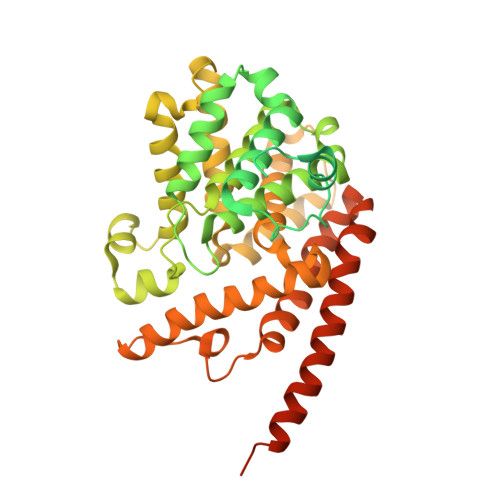
Structural Asymmetry of Phosphodiesterase-9A and a Unique Pocket for Selective Binding of a Potent Enantiomeric Inhibitor.

Huang, M.Shao, Y.Hou, J.Cui, W.Liang, B.Huang, Y.Li, Z.Wu, Y.Zhu, X.Liu, P.Wan, Y.Ke, H.Luo, H.B.

(2015) Mol Pharmacol 88: 836-845

  • DOI: https://doi.org/10.1124/mol.115.099747
  • Primary Citation of Related Structures:  
    4Y86, 4Y87, 4Y8C

  • PubMed Abstract: 

    Phosphodiesterase-9 (PDE9) inhibitors have been studied as potential therapeutics for treatment of central nervous system diseases and diabetes. Here, we report the discovery of a new category of PDE9 inhibitors by rational design on the basis of the crystal structures. The best compound, (S)-6-((1-(4-chlorophenyl)ethyl)amino)-1-cyclopentyl-1,5,6,7-tetrahydro-4H-pyrazolo[3,4-day]pyrimidin-4-one [(S)-C33], has an IC50 value of 11 nM against PDE9 and the racemic C33 has bioavailability of 56.5% in the rat pharmacokinetic model. The crystal structures of PDE9 in the complex with racemic C33, (R)-C33, and (S)-C33 reveal subtle conformational asymmetry of two M-loops in the PDE9 dimer and different conformations of two C33 enantiomers. The structures also identified a small hydrophobic pocket that interacts with the tyrosyl tail of (S)-C33 but not with (R)-C33, and is thus possibly useful for improvement of selectivity of PDE9 inhibitors. The asymmetry of the M-loop and the different interactions of the C33 enantiomers imply the necessity to consider the whole PDE9 dimer in the design of inhibitors.


  • Organizational Affiliation

    School of Chemistry and Chemical Engineering (M.H., J.H., X.Z. Yiq.W.), School of Pharmaceutical Sciences (Y.S., Z.L., Yin.W., P.L., H.-B.L.), Sun Yat-Sen University, Guangzhou, PR China; and Department of Biochemistry and Biophysics and Lineberger Comprehensive Cancer Center, University of North Carolina, Chapel Hill, North Carolina (W.C., B.L., Y.H., H.K.).


Macromolecules
Find similar proteins by:  (by identity cutoff)  |  3D Structure
Entity ID: 1
MoleculeChains Sequence LengthOrganismDetailsImage
High affinity cGMP-specific 3',5'-cyclic phosphodiesterase 9A
A, B
533Homo sapiensMutation(s): 0 
Gene Names: PDE9A
EC: 3.1.4.35
UniProt & NIH Common Fund Data Resources
Find proteins for O76083 (Homo sapiens)
Explore O76083 
Go to UniProtKB:  O76083
PHAROS:  O76083
GTEx:  ENSG00000160191 
Entity Groups  
Sequence Clusters30% Identity50% Identity70% Identity90% Identity95% Identity100% Identity
UniProt GroupO76083
Sequence Annotations
Expand
  • Reference Sequence
Small Molecules
Ligands 4 Unique
IDChains Name / Formula / InChI Key2D Diagram3D Interactions
49E
Query on 49E

Download Ideal Coordinates CCD File 
F [auth B]6-{[(1R)-1-(4-chlorophenyl)ethyl]amino}-1-cyclopentyl-1,5-dihydro-4H-pyrazolo[3,4-d]pyrimidin-4-one
C18 H20 Cl N5 O
FIUCLBJMUGCQTF-LLVKDONJSA-N
49D
Query on 49D

Download Ideal Coordinates CCD File 
C [auth A]6-{[(1S)-1-(4-chlorophenyl)ethyl]amino}-1-cyclopentyl-1,5-dihydro-4H-pyrazolo[3,4-d]pyrimidin-4-one
C18 H20 Cl N5 O
FIUCLBJMUGCQTF-NSHDSACASA-N
ZN
Query on ZN

Download Ideal Coordinates CCD File 
D [auth A],
G [auth B]
ZINC ION
Zn
PTFCDOFLOPIGGS-UHFFFAOYSA-N
MG
Query on MG

Download Ideal Coordinates CCD File 
E [auth A],
H [auth B]
MAGNESIUM ION
Mg
JLVVSXFLKOJNIY-UHFFFAOYSA-N
Binding Affinity Annotations 
IDSourceBinding Affinity
49D BindingDB:  4Y86 IC50: 6 (nM) from 1 assay(s)
Binding MOAD:  4Y86 IC50: 11 (nM) from 1 assay(s)
49E Binding MOAD:  4Y86 IC50: 58 (nM) from 1 assay(s)
Experimental Data & Validation

Experimental Data

  • Method: X-RAY DIFFRACTION
  • Resolution: 2.01 Å
  • R-Value Free: 0.247 
  • R-Value Work: 0.223 
  • R-Value Observed: 0.224 
  • Space Group: P 41 21 2
Unit Cell:
Length ( Å )Angle ( ˚ )
a = 104.135α = 90
b = 104.135β = 90
c = 269.536γ = 90
Software Package:
Software NamePurpose
REFMACrefinement
HKL-2000data reduction
HKL-2000data scaling
AMoREphasing

Structure Validation

View Full Validation Report


Ligand Structure Quality Assessment 


Entry History & Funding Information

Deposition Data

  • Released Date: 2015-09-16 
  • Deposition Author(s): Huang, M.

Funding OrganizationLocationGrant Number
National Institutes of Health/National Institute of General Medical Sciences (NIH/NIGMS)United StatesGM59791

Revision History  (Full details and data files)

  • Version 1.0: 2015-09-16
    Type: Initial release
  • Version 1.1: 2015-10-14
    Changes: Database references
  • Version 1.2: 2017-09-20
    Changes: Author supporting evidence, Database references, Derived calculations
  • Version 1.3: 2019-12-25
    Changes: Author supporting evidence
  • Version 1.4: 2024-02-28
    Changes: Data collection, Database references, Derived calculations