4ZEL

Human dopamine beta-hydroxylase


Experimental Data Snapshot

  • Method: X-RAY DIFFRACTION
  • Resolution: 2.90 Å
  • R-Value Free: 0.270 
  • R-Value Work: 0.230 
  • R-Value Observed: 0.232 

wwPDB Validation   3D Report Full Report


Ligand Structure Quality Assessment 


This is version 2.0 of the entry. See complete history


Literature

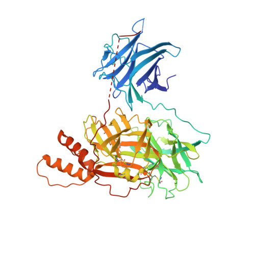
The crystal structure of human dopamine beta-hydroxylase at 2.9 angstrom resolution.

Vendelboe, T.V.Harris, P.Zhao, Y.Walter, T.S.Harlos, K.El Omari, K.Christensen, H.E.

(2016) Sci Adv 2: e1500980-e1500980

  • DOI: https://doi.org/10.1126/sciadv.1500980
  • Primary Citation of Related Structures:  
    4ZEL

  • PubMed Abstract: 

    The norepinephrine pathway is believed to modulate behavioral and physiological processes, such as mood, overall arousal, and attention. Furthermore, abnormalities in the pathway have been linked to numerous diseases, for example hypertension, depression, anxiety, Parkinson's disease, schizophrenia, Alzheimer's disease, attention deficit hyperactivity disorder, and cocaine dependence. We report the crystal structure of human dopamine β-hydroxylase, which is the enzyme converting dopamine to norepinephrine. The structure of the DOMON (dopamine β-monooxygenase N-terminal) domain, also found in >1600 other proteins, reveals a possible metal-binding site and a ligand-binding pocket. The catalytic core structure shows two different conformations: an open active site, as also seen in another member of this enzyme family [the peptidylglycine α-hydroxylating (and α-amidating) monooxygenase], and a closed active site structure, in which the two copper-binding sites are only 4 to 5 Å apart, in what might be a coupled binuclear copper site. The dimerization domain adopts a conformation that bears no resemblance to any other known protein structure. The structure provides new molecular insights into the numerous devastating disorders of both physiological and neurological origins associated with the dopamine system.


  • Organizational Affiliation

    Department of Chemistry, Kemitorvet 207, Technical University of Denmark, DK-2800 Kgs. Lyngby, Denmark.


Macromolecules
Find similar proteins by:  (by identity cutoff)  |  3D Structure
Entity ID: 1
MoleculeChains Sequence LengthOrganismDetailsImage
Dopamine beta-hydroxylase
A, B
578Homo sapiensMutation(s): 0 
Gene Names: DBH
EC: 1.14.17.1
UniProt & NIH Common Fund Data Resources
Find proteins for P09172 (Homo sapiens)
Explore P09172 
Go to UniProtKB:  P09172
PHAROS:  P09172
GTEx:  ENSG00000123454 
Entity Groups  
Sequence Clusters30% Identity50% Identity70% Identity90% Identity95% Identity100% Identity
UniProt GroupP09172
Sequence Annotations
Expand
  • Reference Sequence
Oligosaccharides

Help

Entity ID: 2
MoleculeChains Length2D Diagram Glycosylation3D Interactions
2-acetamido-2-deoxy-beta-D-glucopyranose-(1-4)-2-acetamido-2-deoxy-beta-D-glucopyranose
C
2N-Glycosylation
Glycosylation Resources
GlyTouCan:  G42666HT
GlyCosmos:  G42666HT
GlyGen:  G42666HT
Entity ID: 3
MoleculeChains Length2D Diagram Glycosylation3D Interactions
beta-D-mannopyranose-(1-4)-2-acetamido-2-deoxy-beta-D-glucopyranose-(1-4)-2-acetamido-2-deoxy-beta-D-glucopyranose
D, F
3N-Glycosylation
Glycosylation Resources
GlyTouCan:  G15407YE
GlyCosmos:  G15407YE
GlyGen:  G15407YE
Entity ID: 4
MoleculeChains Length2D Diagram Glycosylation3D Interactions
alpha-D-mannopyranose-(1-6)-beta-D-mannopyranose-(1-4)-2-acetamido-2-deoxy-beta-D-glucopyranose-(1-4)-2-acetamido-2-deoxy-beta-D-glucopyranose
E
4N-Glycosylation
Glycosylation Resources
GlyTouCan:  G22573RC
GlyCosmos:  G22573RC
GlyGen:  G22573RC
Experimental Data & Validation

Experimental Data

  • Method: X-RAY DIFFRACTION
  • Resolution: 2.90 Å
  • R-Value Free: 0.270 
  • R-Value Work: 0.230 
  • R-Value Observed: 0.232 
  • Space Group: C 2 2 21
Unit Cell:
Length ( Å )Angle ( ˚ )
a = 102.81α = 90
b = 119.06β = 90
c = 224.83γ = 90
Software Package:
Software NamePurpose
PHENIXrefinement
xia2data reduction
PDB_EXTRACTdata extraction
xia2data scaling
SHELXDEphasing

Structure Validation

View Full Validation Report


Ligand Structure Quality Assessment 


Entry History 

Deposition Data

Revision History  (Full details and data files)

  • Version 1.0: 2016-04-20
    Type: Initial release
  • Version 1.1: 2016-05-18
    Changes: Database references
  • Version 2.0: 2020-07-29
    Type: Remediation
    Reason: Carbohydrate remediation
    Changes: Atomic model, Data collection, Derived calculations, Structure summary