5DHH | pdb_00005dhh

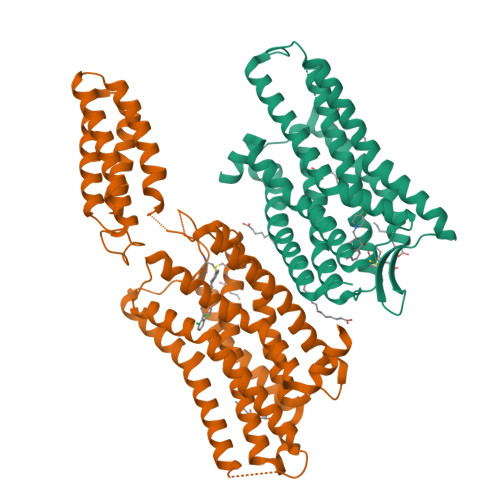
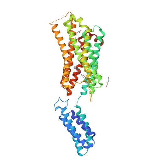
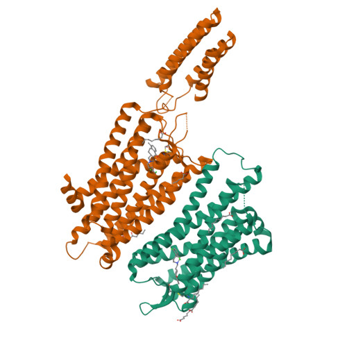
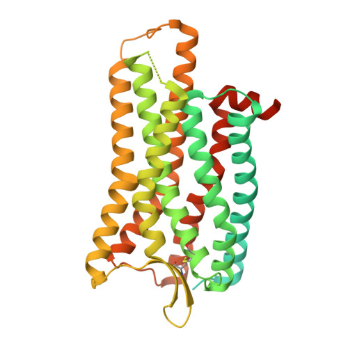
The crystal structure of nociceptin/orphanin FQ peptide receptor (NOP) in complex with SB-612111 (PSI Community Target)


Experimental Data Snapshot

  • Method: X-RAY DIFFRACTION
  • Resolution: 3.00 Å
  • R-Value Free: 
    0.279 (Depositor), 0.280 (DCC) 
  • R-Value Work: 
    0.240 (Depositor), 0.240 (DCC) 
  • R-Value Observed: 
    0.242 (Depositor) 

Starting Model: experimental
View more details

wwPDB Validation   3D Report Full Report


Ligand Structure Quality Assessment 

Created with Raphaël 2.3.0Worse 01 BetterLigand structure goodness of fit to experimental dataBest fitted DGWClick on this verticalbar to view detailsBest fitted OLCClick on this verticalbar to view detailsBest fitted OLAClick on this verticalbar to view details

This is version 1.6 of the entry. See complete history


Literature

The Importance of Ligand-Receptor Conformational Pairs in Stabilization: Spotlight on the N/OFQ G Protein-Coupled Receptor.

Miller, R.L.Thompson, A.A.Trapella, C.Guerrini, R.Malfacini, D.Patel, N.Han, G.W.Cherezov, V.Calo, G.Katritch, V.Stevens, R.C.

(2015) Structure 23: 2291-2299

  • DOI: https://doi.org/10.1016/j.str.2015.07.024
  • Primary Citation of Related Structures:  
    5DHG, 5DHH

  • PubMed Abstract: 

    Understanding the mechanism by which ligands affect receptor conformational equilibria is key in accelerating membrane protein structural biology. In the case of G protein-coupled receptors (GPCRs), we currently pursue a brute-force approach for identifying ligands that stabilize receptors and facilitate crystallogenesis. The nociceptin/orphanin FQ peptide receptor (NOP) is a member of the opioid receptor subfamily of GPCRs for which many structurally diverse ligands are available for screening. We observed that antagonist potency is correlated with a ligand's ability to induce receptor stability (Tm) and crystallogenesis. Using this screening strategy, we solved two structures of NOP in complex with top candidate ligands SB-612111 and C-35. Docking studies indicate that while potent, stabilizing antagonists strongly favor a single binding orientation, less potent ligands can adopt multiple binding modes, contributing to their low Tm values. These results suggest a mechanism for ligand-aided crystallogenesis whereby potent antagonists stabilize a single ligand-receptor conformational pair.


  • Organizational Affiliation

    Department of Integrative Structural and Computational Biology, The Scripps Research Institute, La Jolla, CA 92037, USA.


Macromolecules
Find similar proteins by:  (by identity cutoff)  |  3D Structure
Entity ID: 1
MoleculeChains Sequence LengthOrganismDetailsImage
Soluble cytochrome b562,Nociceptin receptor
A, B
424Escherichia coliHomo sapiens
This entity is chimeric
Mutation(s): 3 
Gene Names: cybCOPRL1OORORL1
Membrane Entity: Yes 
UniProt & NIH Common Fund Data Resources
Find proteins for P41146 (Homo sapiens)
Explore P41146 
Go to UniProtKB:  P41146
PHAROS:  P41146
GTEx:  ENSG00000125510 
Find proteins for P0ABE7 (Escherichia coli)
Explore P0ABE7 
Go to UniProtKB:  P0ABE7
Entity Groups  
Sequence Clusters30% Identity50% Identity70% Identity90% Identity95% Identity100% Identity
UniProt GroupsP41146P0ABE7
Sequence Annotations
Expand
  • Reference Sequence
Small Molecules
Ligands 3 Unique
IDChains Name / Formula / InChI Key2D Diagram3D Interactions
DGW
Query on DGW

Download Ideal Coordinates CCD File 
H [auth A],
K [auth B]
(5S,7S)-7-{[4-(2,6-dichlorophenyl)piperidin-1-yl]methyl}-1-methyl-6,7,8,9-tetrahydro-5H-benzo[7]annulen-5-ol
C24 H29 Cl2 N O
OHRDCQFCAWLDBP-SBUREZEXSA-N
OLC
Query on OLC

Download Ideal Coordinates CCD File 
L [auth B](2R)-2,3-dihydroxypropyl (9Z)-octadec-9-enoate
C21 H40 O4
RZRNAYUHWVFMIP-GDCKJWNLSA-N
OLA
Query on OLA

Download Ideal Coordinates CCD File 
C [auth A]
D [auth A]
E [auth A]
F [auth A]
G [auth A]
OLEIC ACID
C18 H34 O2
ZQPPMHVWECSIRJ-KTKRTIGZSA-N
Binding Affinity Annotations 
IDSourceBinding Affinity
DGW BindingDB:  5DHH Ki: min: 0.25, max: 0.33 (nM) from 2 assay(s)
Experimental Data & Validation

Experimental Data

  • Method: X-RAY DIFFRACTION
  • Resolution: 3.00 Å
  • R-Value Free:  0.279 (Depositor), 0.280 (DCC) 
  • R-Value Work:  0.240 (Depositor), 0.240 (DCC) 
  • R-Value Observed: 0.242 (Depositor) 
Space Group: P 1 21 1
Unit Cell:
Length ( Å )Angle ( ˚ )
a = 42.3α = 90
b = 168.941β = 103.5
c = 65.472γ = 90
Software Package:
Software NamePurpose
PHENIXrefinement
DENZOdata reduction
SCALEPACKdata scaling
PHASERphasing

Structure Validation

View Full Validation Report


Ligand Structure Quality Assessment 

Created with Raphaël 2.3.0Worse 01 BetterLigand structure goodness of fit to experimental dataBest fitted DGWClick on this verticalbar to view detailsBest fitted OLCClick on this verticalbar to view detailsBest fitted OLAClick on this verticalbar to view details

Entry History 

Revision History  (Full details and data files)

  • Version 1.0: 2015-10-21
    Type: Initial release
  • Version 1.1: 2015-11-18
    Changes: Database references
  • Version 1.2: 2015-12-16
    Changes: Database references
  • Version 1.3: 2017-05-31
    Changes: Source and taxonomy
  • Version 1.4: 2017-07-05
    Changes: Database references
  • Version 1.5: 2023-09-27
    Changes: Data collection, Database references, Refinement description
  • Version 1.6: 2024-10-23
    Changes: Structure summary