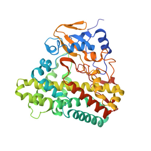
5EDT

Crystal structure of Mycobacterium tuberculosis CYP121 in complex with LIG9


Experimental Data Snapshot

  • Method: X-RAY DIFFRACTION
  • Resolution: 2.45 Å
  • R-Value Free: 0.243 
  • R-Value Work: 0.187 
  • R-Value Observed: 0.190 

Starting Model: experimental
View more details

wwPDB Validation   3D Report Full Report


Ligand Structure Quality Assessment 


This is version 2.2 of the entry. See complete history


Literature

Fragment-Based Approaches to the Development of Mycobacterium tuberculosis CYP121 Inhibitors.

Kavanagh, M.E.Coyne, A.G.McLean, K.J.James, G.G.Levy, C.W.Marino, L.B.de Carvalho, L.P.Chan, D.S.Hudson, S.A.Surade, S.Leys, D.Munro, A.W.Abell, C.

(2016) J Med Chem 59: 3272-3302

  • DOI: https://doi.org/10.1021/acs.jmedchem.6b00007
  • Primary Citation of Related Structures:  
    5EDT, 5IBD, 5IBE, 5IBF, 5IBG, 5IBH, 5IBI, 5IBJ

  • PubMed Abstract: 

    The essential enzyme CYP121 is a target for drug development against antibiotic resistant strains of Mycobacterium tuberculosis. A triazol-1-yl phenol fragment 1 was identified to bind to CYP121 using a cascade of biophysical assays. Synthetic merging and optimization of 1 produced a 100-fold improvement in binding affinity, yielding lead compound 2 (KD = 15 μM). Deconstruction of 2 into its component retrofragments allowed the group efficiency of structural motifs to be assessed, the identification of more LE scaffolds for optimization and highlighted binding affinity hotspots. Structure-guided addition of a metal-binding pharmacophore onto LE retrofragment scaffolds produced low nanomolar (KD = 15 nM) CYP121 ligands. Elaboration of these compounds to target binding hotspots in the distal active site afforded compounds with excellent selectivity against human drug-metabolizing P450s. Analysis of the factors governing ligand potency and selectivity using X-ray crystallography, UV-vis spectroscopy, and native mass spectrometry provides insight for subsequent drug development.


  • Organizational Affiliation

    Department of Chemistry, University of Cambridge , Lensfield Road, Cambridge CB2 1EW, U.K.


Macromolecules
Find similar proteins by:  (by identity cutoff)  |  3D Structure
Entity ID: 1
MoleculeChains Sequence LengthOrganismDetailsImage
Cytochrome P450 121395Mycobacterium tuberculosis H37RvMutation(s): 0 
Gene Names: cyp121Mb2299
EC: 1.14
UniProt
Find proteins for P0A515 (Mycobacterium bovis (strain ATCC BAA-935 / AF2122/97))
Explore P0A515 
Go to UniProtKB:  P0A515
Entity Groups  
Sequence Clusters30% Identity50% Identity70% Identity90% Identity95% Identity100% Identity
UniProt GroupP0A515
Sequence Annotations
Expand
  • Reference Sequence
Small Molecules
Ligands 3 Unique
IDChains Name / Formula / InChI Key2D Diagram3D Interactions
HEM
Query on HEM

Download Ideal Coordinates CCD File 
B [auth A]PROTOPORPHYRIN IX CONTAINING FE
C34 H32 Fe N4 O4
KABFMIBPWCXCRK-RGGAHWMASA-L
5MK
Query on 5MK

Download Ideal Coordinates CCD File 
C [auth A]2-azanyl-5-chloranyl-benzamide
C7 H7 Cl N2 O
DNRVZOZGQHHDAT-UHFFFAOYSA-N
SO4
Query on SO4

Download Ideal Coordinates CCD File 
D [auth A]
E [auth A]
F [auth A]
G [auth A]
H [auth A]
D [auth A],
E [auth A],
F [auth A],
G [auth A],
H [auth A],
I [auth A],
J [auth A],
K [auth A],
L [auth A],
M [auth A]
SULFATE ION
O4 S
QAOWNCQODCNURD-UHFFFAOYSA-L
Experimental Data & Validation

Experimental Data

  • Method: X-RAY DIFFRACTION
  • Resolution: 2.45 Å
  • R-Value Free: 0.243 
  • R-Value Work: 0.187 
  • R-Value Observed: 0.190 
  • Space Group: P 65 2 2
Unit Cell:
Length ( Å )Angle ( ˚ )
a = 77α = 90
b = 77β = 90
c = 263γ = 120
Software Package:
Software NamePurpose
REFMACrefinement
XPREPdata scaling
PHASERphasing

Structure Validation

View Full Validation Report


Ligand Structure Quality Assessment 


Entry History & Funding Information

Deposition Data


Funding OrganizationLocationGrant Number
Biotechnology and Biological Sciences Research CouncilUnited KingdomBB/I019669/1
University of CambridgeUnited KingdomCambridge Commonwealth Trust and Cambridge Overseas Trust

Revision History  (Full details and data files)

  • Version 1.0: 2016-04-06
    Type: Initial release
  • Version 1.1: 2016-04-27
    Changes: Database references
  • Version 2.0: 2017-08-30
    Changes: Atomic model, Author supporting evidence
  • Version 2.1: 2018-11-21
    Changes: Data collection, Source and taxonomy
  • Version 2.2: 2024-01-10
    Changes: Data collection, Database references, Refinement description