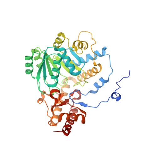
5LUC

Crystal structure of the D183N variant of human Alanine:Glyoxylate Aminotransferase major allele (AGT-Ma) at 1.8 Angstrom; internal aldimine with PLP in the active site


Experimental Data Snapshot

  • Method: X-RAY DIFFRACTION
  • Resolution: 1.80 Å
  • R-Value Free: 0.192 
  • R-Value Work: 0.163 
  • R-Value Observed: 0.165 

Starting Model: experimental
View more details

wwPDB Validation   3D Report Full Report


Ligand Structure Quality Assessment 


This is version 1.2 of the entry. See complete history


Literature

Radiation damage at the active site of human alanine:glyoxylate aminotransferase reveals that the cofactor position is finely tuned during catalysis.

Giardina, G.Paiardini, A.Montioli, R.Cellini, B.Voltattorni, C.B.Cutruzzola, F.

(2017) Sci Rep 7: 11704-11704

  • DOI: https://doi.org/10.1038/s41598-017-11948-w
  • Primary Citation of Related Structures:  
    5F9S, 5HHY, 5LUC, 5OFY, 5OG0

  • PubMed Abstract: 

    The alanine:glyoxylate aminotransferase (AGT), a hepatocyte-specific pyridoxal-5'-phosphate (PLP) dependent enzyme, transaminates L-alanine and glyoxylate to glycine and pyruvate, thus detoxifying glyoxylate and preventing pathological oxalate precipitation in tissues. In the widely accepted catalytic mechanism of the aminotransferase family, the lysine binding to PLP acts as a catalyst in the stepwise 1,3-proton transfer, interconverting the external aldimine to ketimine. This step requires protonation by a conserved aspartate of the pyridine nitrogen of PLP to enhance its ability to stabilize the carbanionic intermediate. The aspartate residue is also responsible for a significant geometrical distortion of the internal aldimine, crucial for catalysis. We present the structure of human AGT in which complete X-ray photoreduction of the Schiff base has occurred. This result, together with two crystal structures of the conserved aspartate pathogenic variant (D183N) and the molecular modeling of the transaldimination step, led us to propose that an interplay of opposite forces, which we named spring mechanism, finely tunes PLP geometry during catalysis and is essential to move the external aldimine in the correct position in order for the 1,3-proton transfer to occur.


  • Organizational Affiliation

    Department of Biochemical Sciences "A. Rossi Fanelli", Sapienza University of Rome, Rome, Italy. giorgio.giardina@uniroma1.it.


Macromolecules
Find similar proteins by:  (by identity cutoff)  |  3D Structure
Entity ID: 1
MoleculeChains Sequence LengthOrganismDetailsImage
Serine--pyruvate aminotransferase403Homo sapiensMutation(s): 1 
Gene Names: AGXTAGT1SPAT
EC: 2.6.1.51 (PDB Primary Data), 2.6.1.44 (PDB Primary Data)
UniProt & NIH Common Fund Data Resources
Find proteins for P21549 (Homo sapiens)
Explore P21549 
Go to UniProtKB:  P21549
PHAROS:  P21549
GTEx:  ENSG00000172482 
Entity Groups  
Sequence Clusters30% Identity50% Identity70% Identity90% Identity95% Identity100% Identity
UniProt GroupP21549
Sequence Annotations
Expand
  • Reference Sequence
Small Molecules
Ligands 2 Unique
IDChains Name / Formula / InChI Key2D Diagram3D Interactions
PLP
Query on PLP

Download Ideal Coordinates CCD File 
I [auth A]
K [auth B]
M [auth E]
O [auth G]
Q [auth M]
I [auth A],
K [auth B],
M [auth E],
O [auth G],
Q [auth M],
S [auth N],
U [auth S],
W [auth T]
PYRIDOXAL-5'-PHOSPHATE
C8 H10 N O6 P
NGVDGCNFYWLIFO-UHFFFAOYSA-N
BTB
Query on BTB

Download Ideal Coordinates CCD File 
J [auth A]
L [auth B]
N [auth E]
P [auth G]
R [auth M]
J [auth A],
L [auth B],
N [auth E],
P [auth G],
R [auth M],
T [auth N],
V [auth S],
X [auth T]
2-[BIS-(2-HYDROXY-ETHYL)-AMINO]-2-HYDROXYMETHYL-PROPANE-1,3-DIOL
C8 H19 N O5
OWMVSZAMULFTJU-UHFFFAOYSA-N
Experimental Data & Validation

Experimental Data

  • Method: X-RAY DIFFRACTION
  • Resolution: 1.80 Å
  • R-Value Free: 0.192 
  • R-Value Work: 0.163 
  • R-Value Observed: 0.165 
  • Space Group: P 21 21 21
Unit Cell:
Length ( Å )Angle ( ˚ )
a = 127.599α = 90
b = 141.107β = 90
c = 256.117γ = 90
Software Package:
Software NamePurpose
Aimlessdata scaling
MOLREPphasing
PHENIXrefinement
PDB_EXTRACTdata extraction
XDSdata reduction
MOLREPphasing

Structure Validation

View Full Validation Report


Ligand Structure Quality Assessment 


Entry History 

Deposition Data

Revision History  (Full details and data files)

  • Version 1.0: 2017-09-20
    Type: Initial release
  • Version 1.1: 2017-09-27
    Changes: Database references
  • Version 1.2: 2024-01-17
    Changes: Advisory, Data collection, Database references, Refinement description