5UAN

Crystal structure of multi-domain RAR-beta-RXR-alpha heterodimer on DNA


Experimental Data Snapshot

  • Method: X-RAY DIFFRACTION
  • Resolution: 3.51 Å
  • R-Value Free: 0.270 
  • R-Value Work: 0.221 
  • R-Value Observed: 0.223 

wwPDB Validation   3D Report Full Report


Ligand Structure Quality Assessment 


This is version 1.2 of the entry. See complete history


Literature

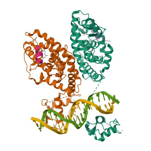
The quaternary architecture of RAR beta-RXR alpha heterodimer facilitates domain-domain signal transmission.

Chandra, V.Wu, D.Li, S.Potluri, N.Kim, Y.Rastinejad, F.

(2017) Nat Commun 8: 868-868

  • DOI: https://doi.org/10.1038/s41467-017-00981-y
  • Primary Citation of Related Structures:  
    5UAN

  • PubMed Abstract: 

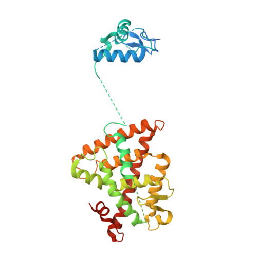
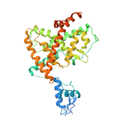
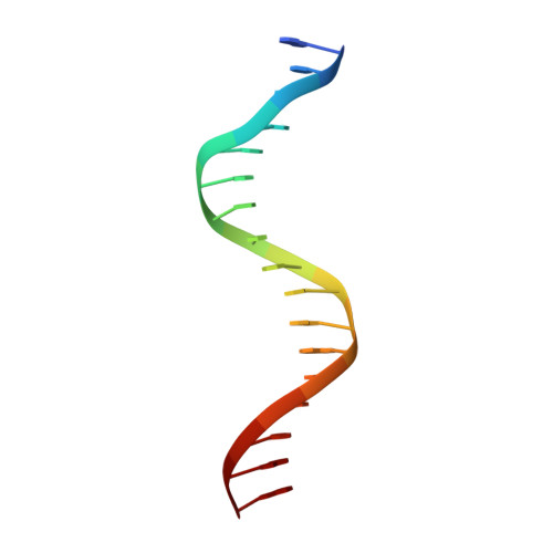
    Assessing the physical connections and allosteric communications in multi-domain nuclear receptor (NR) polypeptides has remained challenging, with few crystal structures available to show their overall structural organizations. Here we report the quaternary architecture of multi-domain retinoic acid receptor β-retinoic X receptor α (RARβ-RXRα) heterodimer bound to DNA, ligands and coactivator peptides, examined through crystallographic, hydrogen-deuterium exchange mass spectrometry, mutagenesis and functional studies. The RARβ ligand-binding domain (LBD) and DNA-binding domain (DBD) are physically connected to foster allosteric signal transmission between them. Direct comparisons among all the multi-domain NRs studied crystallographically to date show significant variations within their quaternary architectures, rather than a common architecture adhering to strict rules. RXR remains flexible and adaptive by maintaining loosely organized domains, while its heterodimerization partners use a surface patch on their LBDs to form domain-domain interactions with DBDs.Nuclear receptors (NR) are multidomain proteins, which makes their crystallization challenging. Here the authors present the crystal structure of the retinoic acid receptor β-retinoic X receptor α (RARβ-RXRα) heterodimer bound to DNA, ligands and coactivator peptides, which shows that NR quaternary architectures are variable.


  • Organizational Affiliation

    Integrative Metabolism Program, Sanford Burnham Prebys Medical Discovery Institute, Orlando, FL, 32827, USA.


Macromolecules

Find similar proteins by:  (by identity cutoff)  |  3D Structure
Entity ID: 1
MoleculeChains Sequence LengthOrganismDetailsImage
Retinoic acid receptor RXR-alpha365Homo sapiensMutation(s): 0 
Gene Names: RXRANR2B1
UniProt & NIH Common Fund Data Resources
Find proteins for P19793 (Homo sapiens)
Explore P19793 
Go to UniProtKB:  P19793
PHAROS:  P19793
GTEx:  ENSG00000186350 
Entity Groups  
Sequence Clusters30% Identity50% Identity70% Identity90% Identity95% Identity100% Identity
UniProt GroupP19793
Sequence Annotations
Expand
  • Reference Sequence
Find similar proteins by:  (by identity cutoff)  |  3D Structure
Entity ID: 2
MoleculeChains Sequence LengthOrganismDetailsImage
Retinoic acid receptor beta397Homo sapiensMutation(s): 0 
Gene Names: RARBHAPNR1B2
UniProt & NIH Common Fund Data Resources
Find proteins for P10826 (Homo sapiens)
Explore P10826 
Go to UniProtKB:  P10826
PHAROS:  P10826
GTEx:  ENSG00000077092 
Entity Groups  
Sequence Clusters30% Identity50% Identity70% Identity90% Identity95% Identity100% Identity
UniProt GroupP10826
Sequence Annotations
Expand
  • Reference Sequence

Find similar proteins by:  Sequence   |   3D Structure  

Entity ID: 3
MoleculeChains Sequence LengthOrganismDetailsImage
Nuclear receptor coactivator 2
C, D
10Homo sapiensMutation(s): 0 
UniProt & NIH Common Fund Data Resources
Find proteins for Q15596 (Homo sapiens)
Explore Q15596 
Go to UniProtKB:  Q15596
PHAROS:  Q15596
GTEx:  ENSG00000140396 
Entity Groups  
Sequence Clusters30% Identity50% Identity70% Identity90% Identity95% Identity100% Identity
UniProt GroupQ15596
Sequence Annotations
Expand
  • Reference Sequence

Find similar nucleic acids by:  Sequence   |   3D Structure  

Entity ID: 4
MoleculeChains LengthOrganismImage
DNA (5'-D(*CP*TP*AP*GP*GP*TP*CP*AP*AP*AP*GP*GP*TP*CP*AP*GP*C)-3')17Homo sapiens
Sequence Annotations
Expand
  • Reference Sequence

Find similar nucleic acids by:  Sequence   |   3D Structure  

Entity ID: 5
MoleculeChains LengthOrganismImage
DNA (5'-D(*GP*CP*TP*GP*AP*CP*CP*TP*TP*TP*GP*AP*CP*CP*TP*AP*G)-3')17Homo sapiens
Sequence Annotations
Expand
  • Reference Sequence
Small Molecules
Binding Affinity Annotations 
IDSourceBinding Affinity
9CR BindingDB:  5UAN Ki: min: 0.5, max: 583 (nM) from 11 assay(s)
Kd: min: 0.8, max: 1810 (nM) from 24 assay(s)
IC50: min: 7, max: 82 (nM) from 7 assay(s)
EC50: min: 1.5, max: 316 (nM) from 32 assay(s)
REA BindingDB:  5UAN IC50: min: 1000, max: 5.00e+4 (nM) from 4 assay(s)
Experimental Data & Validation

Experimental Data

  • Method: X-RAY DIFFRACTION
  • Resolution: 3.51 Å
  • R-Value Free: 0.270 
  • R-Value Work: 0.221 
  • R-Value Observed: 0.223 
  • Space Group: P 1 21 1
Unit Cell:
Length ( Å )Angle ( ˚ )
a = 52.47α = 90
b = 77.403β = 90.37
c = 112.113γ = 90
Software Package:
Software NamePurpose
HKL-3000data reduction
HKL-3000data scaling
PHASERphasing
PHENIXrefinement
Cootmodel building
PDB_EXTRACTdata extraction

Structure Validation

View Full Validation Report


Ligand Structure Quality Assessment 


Entry History 

Deposition Data

Revision History  (Full details and data files)

  • Version 1.0: 2017-10-18
    Type: Initial release
  • Version 1.1: 2018-04-18
    Changes: Data collection, Database references
  • Version 1.2: 2023-10-04
    Changes: Data collection, Database references, Refinement description