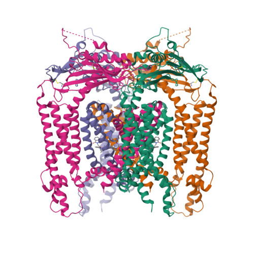
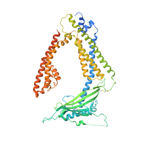
5WJ9

Human TRPML1 channel structure in agonist-bound open conformation


Experimental Data Snapshot

  • Method: ELECTRON MICROSCOPY
  • Resolution: 3.49 Å
  • Aggregation State: PARTICLE 
  • Reconstruction Method: SINGLE PARTICLE 

wwPDB Validation   3D Report Full Report


This is version 1.6 of the entry. See complete history


Literature

Human TRPML1 channel structures in open and closed conformations.

Schmiege, P.Fine, M.Blobel, G.Li, X.

(2017) Nature 550: 366-370

  • DOI: https://doi.org/10.1038/nature24036
  • Primary Citation of Related Structures:  
    5WJ5, 5WJ9

  • PubMed Abstract: 

    Transient receptor potential mucolipin 1 (TRPML1) is a Ca 2+ -releasing cation channel that mediates the calcium signalling and homeostasis of lysosomes. Mutations in TRPML1 lead to mucolipidosis type IV, a severe lysosomal storage disorder. Here we report two electron cryo-microscopy structures of full-length human TRPML1: a 3.72-Å apo structure at pH 7.0 in the closed state, and a 3.49-Å agonist-bound structure at pH 6.0 in an open state. Several aromatic and hydrophobic residues in pore helix 1, helices S5 and S6, and helix S6 of a neighbouring subunit, form a hydrophobic cavity to house the agonist, suggesting a distinct agonist-binding site from that found in TRPV1, a TRP channel from a different subfamily. The opening of TRPML1 is associated with distinct dilations of its lower gate together with a slight structural movement of pore helix 1. Our work reveals the regulatory mechanism of TRPML channels, facilitates better understanding of TRP channel activation, and provides insights into the molecular basis of mucolipidosis type IV pathogenesis.


  • Organizational Affiliation

    Laboratory of Cell Biology, Howard Hughes Medical Institute, The Rockefeller University, New York, New York 10065, USA.


Macromolecules
Find similar proteins by:  (by identity cutoff)  |  3D Structure
Entity ID: 1
MoleculeChains Sequence LengthOrganismDetailsImage
Mucolipin-1
A, B, C, D
580Homo sapiensMutation(s): 0 
Gene Names: MCOLN1ML4MSTP080
Membrane Entity: Yes 
UniProt & NIH Common Fund Data Resources
Find proteins for Q9GZU1 (Homo sapiens)
Explore Q9GZU1 
Go to UniProtKB:  Q9GZU1
PHAROS:  Q9GZU1
GTEx:  ENSG00000090674 
Entity Groups  
Sequence Clusters30% Identity50% Identity70% Identity90% Identity95% Identity100% Identity
UniProt GroupQ9GZU1
Sequence Annotations
Expand
  • Reference Sequence
Experimental Data & Validation

Experimental Data

  • Method: ELECTRON MICROSCOPY
  • Resolution: 3.49 Å
  • Aggregation State: PARTICLE 
  • Reconstruction Method: SINGLE PARTICLE 
EM Software:
TaskSoftware PackageVersion
RECONSTRUCTIONFREALIGN

Structure Validation

View Full Validation Report


Entry History 

Deposition Data

Revision History  (Full details and data files)

  • Version 1.0: 2017-10-18
    Type: Initial release
  • Version 1.1: 2017-10-25
    Changes: Data processing, Database references, Structure summary
  • Version 1.2: 2017-11-01
    Changes: Database references, Structure summary
  • Version 1.3: 2017-11-08
    Changes: Derived calculations
  • Version 1.4: 2018-10-03
    Changes: Data collection, Other, Refinement description
  • Version 1.5: 2019-12-18
    Changes: Other
  • Version 1.6: 2024-11-06
    Changes: Data collection, Database references, Refinement description, Structure summary