6CZS

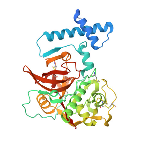
Crystal structure of human pro-cathepsin H C26S mutant


Experimental Data Snapshot

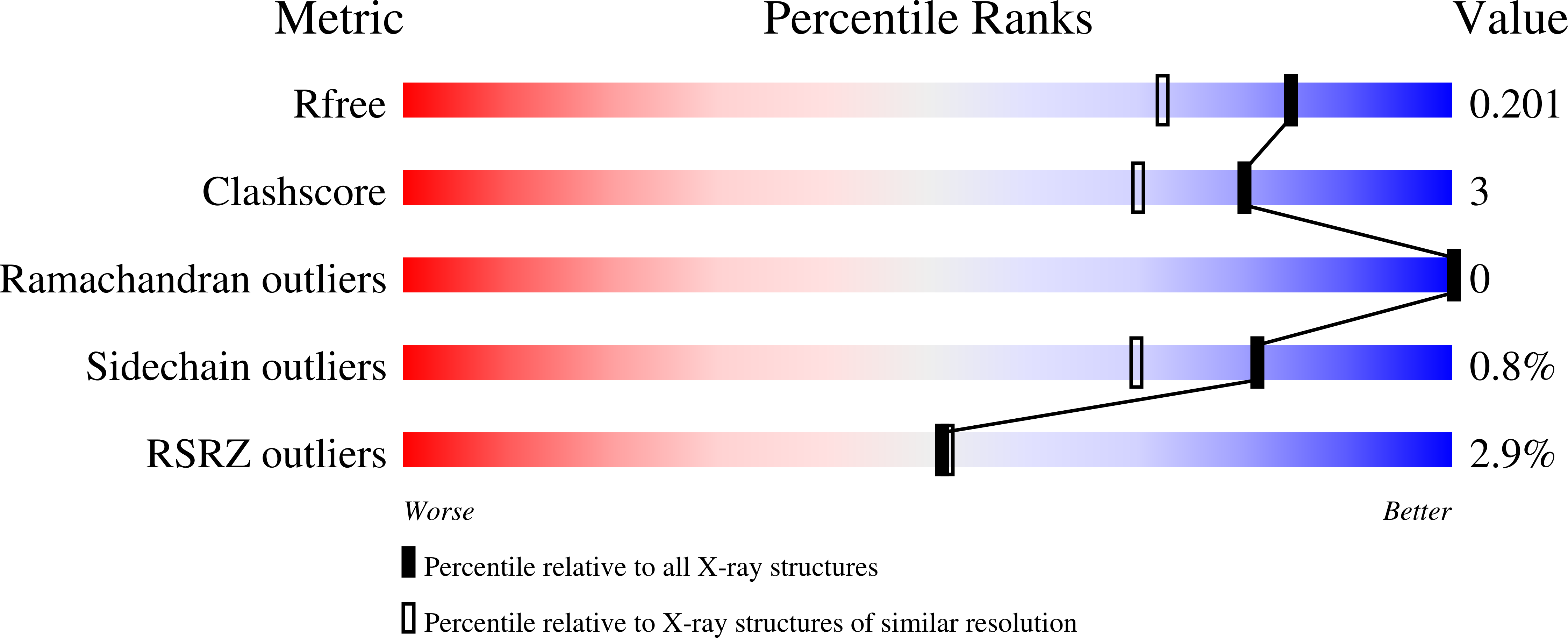
  • Method: X-RAY DIFFRACTION
  • Resolution: 1.66 Å
  • R-Value Free: 0.208 
  • R-Value Work: 0.181 
  • R-Value Observed: 0.182 

Starting Model: experimental
View more details

wwPDB Validation   3D Report Full Report


Ligand Structure Quality Assessment 


This is version 2.2 of the entry. See complete history


Literature

Crystal structures of human procathepsin H.

Hao, Y.Purtha, W.Cortesio, C.Rui, H.Gu, Y.Chen, H.Sickmier, E.A.Manzanillo, P.Huang, X.

(2018) PLoS One 13: e0200374-e0200374

  • DOI: https://doi.org/10.1371/journal.pone.0200374
  • Primary Citation of Related Structures:  
    6CZK, 6CZS

  • PubMed Abstract: 

    Cathepsin H is a member of the papain superfamily of lysosomal cysteine proteases. It is the only known aminopeptidase in the family and is reported to be involved in cancer and other major diseases. Like many other proteases, it is synthesized as an inactive proenzyme. Although the crystal structure of mature porcine cathepsin H revealed the binding of the mini-chain and provided structural basis for the aminopeptidase activity, detailed structural and functional information on the inhibition and activation of procathepsin H has been elusive. Here we present the crystal structures of human procathepsin H at 2.00 Å and 1.66 Å resolution. These structures allow us to explore in detail the molecular basis for the inhibition of the mature domain by the prodomain. Comparison with cathepsin H structure reveals how mini-chain reorients upon activation. We further demonstrate that procathepsin H is not auto-activated but can be trans-activated by cathepsin L.


  • Organizational Affiliation

    Department of Molecular Engineering, Amgen Inc., Cambridge, MA, United States of America.


Macromolecules
Find similar proteins by:  (by identity cutoff)  |  3D Structure
Entity ID: 1
MoleculeChains Sequence LengthOrganismDetailsImage
Pro-cathepsin H335Homo sapiensMutation(s): 1 
Gene Names: CTSHCPSB
EC: 3.4.22.16
UniProt & NIH Common Fund Data Resources
Find proteins for P09668 (Homo sapiens)
Explore P09668 
Go to UniProtKB:  P09668
PHAROS:  P09668
GTEx:  ENSG00000103811 
Entity Groups  
Sequence Clusters30% Identity50% Identity70% Identity90% Identity95% Identity100% Identity
UniProt GroupP09668
Glycosylation
Glycosylation Sites: 2Go to GlyGen: P09668-1
Sequence Annotations
Expand
  • Reference Sequence
Oligosaccharides

Help

Entity ID: 2
MoleculeChains Length2D Diagram Glycosylation3D Interactions
alpha-D-mannopyranose-(1-3)-[alpha-D-mannopyranose-(1-6)]beta-D-mannopyranose-(1-4)-2-acetamido-2-deoxy-beta-D-glucopyranose-(1-4)-2-acetamido-2-deoxy-beta-D-glucopyranose
B
5N-Glycosylation
Glycosylation Resources
GlyTouCan:  G22768VO
GlyCosmos:  G22768VO
GlyGen:  G22768VO
Entity ID: 3
MoleculeChains Length2D Diagram Glycosylation3D Interactions
alpha-D-glucopyranose-(1-1)-alpha-D-glucopyranose
C, D
2N/A
Glycosylation Resources
GlyTouCan:  G92130SN
GlyCosmos:  G92130SN
Small Molecules
Ligands 3 Unique
IDChains Name / Formula / InChI Key2D Diagram3D Interactions
NAG
Query on NAG

Download Ideal Coordinates CCD File 
E [auth A]2-acetamido-2-deoxy-beta-D-glucopyranose
C8 H15 N O6
OVRNDRQMDRJTHS-FMDGEEDCSA-N
SO4
Query on SO4

Download Ideal Coordinates CCD File 
F [auth A],
G [auth A],
H [auth A],
I [auth A],
J [auth A]
SULFATE ION
O4 S
QAOWNCQODCNURD-UHFFFAOYSA-L
CL
Query on CL

Download Ideal Coordinates CCD File 
K [auth A],
L [auth A]
CHLORIDE ION
Cl
VEXZGXHMUGYJMC-UHFFFAOYSA-M
Biologically Interesting Molecules (External Reference) 1 Unique
Experimental Data & Validation

Experimental Data

  • Method: X-RAY DIFFRACTION
  • Resolution: 1.66 Å
  • R-Value Free: 0.208 
  • R-Value Work: 0.181 
  • R-Value Observed: 0.182 
  • Space Group: P 31 2 1
Unit Cell:
Length ( Å )Angle ( ˚ )
a = 98.242α = 90
b = 98.242β = 90
c = 106.586γ = 120
Software Package:
Software NamePurpose
PHENIXrefinement
XDSdata reduction
Aimlessdata scaling
PHASERphasing

Structure Validation

View Full Validation Report


Ligand Structure Quality Assessment 


Entry History 

Deposition Data

Revision History  (Full details and data files)

  • Version 1.0: 2018-08-08
    Type: Initial release
  • Version 2.0: 2020-07-29
    Type: Remediation
    Reason: Carbohydrate remediation
    Changes: Atomic model, Data collection, Derived calculations, Non-polymer description, Structure summary
  • Version 2.1: 2023-10-04
    Changes: Data collection, Database references, Derived calculations, Refinement description, Structure summary
  • Version 2.2: 2024-10-30
    Changes: Structure summary