6HUJ

CryoEM structure of human full-length heteromeric alpha1beta3gamma2L GABA(A)R in complex with picrotoxin, GABA and megabody Mb38.


Experimental Data Snapshot

  • Method: ELECTRON MICROSCOPY
  • Resolution: 3.04 Å
  • Aggregation State: PARTICLE 
  • Reconstruction Method: SINGLE PARTICLE 

wwPDB Validation   3D Report Full Report


This is version 3.0 of the entry. See complete history


Literature

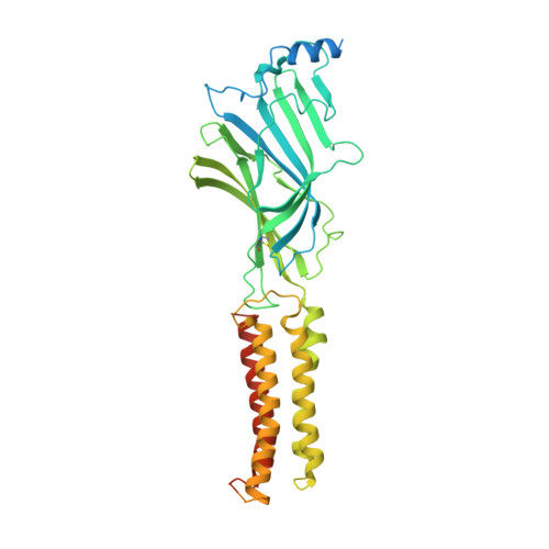
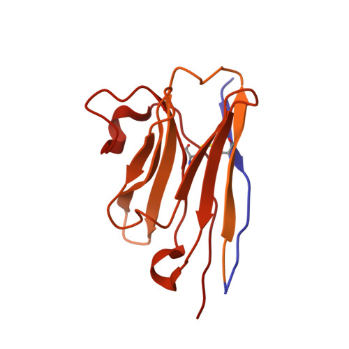
GABAAreceptor signalling mechanisms revealed by structural pharmacology.

Masiulis, S.Desai, R.Uchanski, T.Serna Martin, I.Laverty, D.Karia, D.Malinauskas, T.Zivanov, J.Pardon, E.Kotecha, A.Steyaert, J.Miller, K.W.Aricescu, A.R.

(2019) Nature 565: 454-459

  • DOI: https://doi.org/10.1038/s41586-018-0832-5
  • Primary Citation of Related Structures:  
    6HUG, 6HUJ, 6HUK, 6HUO, 6HUP

  • PubMed Abstract: 

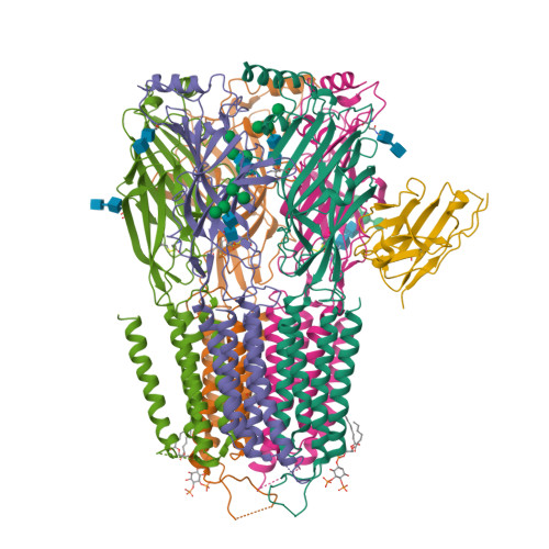
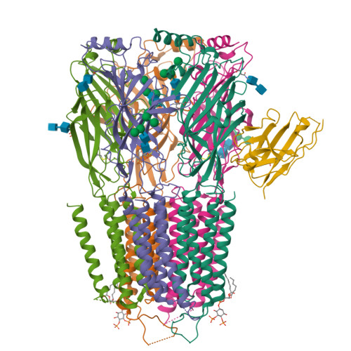
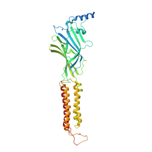
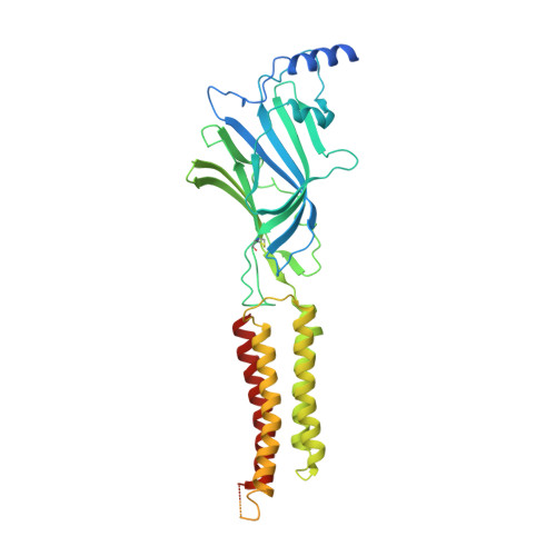
    Type-A γ-aminobutyric (GABA A ) receptors are ligand-gated chloride channels with a very rich pharmacology. Some of their modulators, including benzodiazepines and general anaesthetics, are among the most successful drugs in clinical use and are common substances of abuse. Without reliable structural data, the mechanistic basis for the pharmacological modulation of GABA A receptors remains largely unknown. Here we report several high-resolution cryo-electron microscopy structures in which the full-length human α1β3γ2L GABA A receptor in lipid nanodiscs is bound to the channel-blocker picrotoxin, the competitive antagonist bicuculline, the agonist GABA (γ-aminobutyric acid), and the classical benzodiazepines alprazolam and diazepam. We describe the binding modes and mechanistic effects of these ligands, the closed and desensitized states of the GABA A receptor gating cycle, and the basis for allosteric coupling between the extracellular, agonist-binding region and the transmembrane, pore-forming region. This work provides a structural framework in which to integrate previous physiology and pharmacology research and a rational basis for the development of GABA A receptor modulators.


  • Organizational Affiliation

    MRC Laboratory of Molecular Biology, Cambridge Biomedical Campus, Cambridge, UK. simonasm@mrc-lmb.cam.ac.uk.


Macromolecules
Find similar proteins by:  (by identity cutoff)  |  3D Structure
Entity ID: 1
MoleculeChains Sequence LengthOrganismDetailsImage
Gamma-aminobutyric acid receptor subunit alpha-1,Gamma-aminobutyric acid receptor subunit alpha-1
A, D
464Bos taurusHomo sapiens
This entity is chimeric
Mutation(s): 0 
Gene Names: GABRA1
Membrane Entity: Yes 
UniProt & NIH Common Fund Data Resources
Find proteins for P08219 (Bos taurus)
Explore P08219 
Go to UniProtKB:  P08219
Find proteins for P14867 (Homo sapiens)
Explore P14867 
Go to UniProtKB:  P14867
PHAROS:  P14867
GTEx:  ENSG00000022355 
Entity Groups  
Sequence Clusters30% Identity50% Identity70% Identity90% Identity95% Identity100% Identity
UniProt GroupsP14867P08219
Sequence Annotations
Expand
  • Reference Sequence
Find similar proteins by:  (by identity cutoff)  |  3D Structure
Entity ID: 2
MoleculeChains Sequence LengthOrganismDetailsImage
Gamma-aminobutyric acid receptor subunit beta-3
B, E
473Homo sapiensMutation(s): 0 
Gene Names: GABRB3
Membrane Entity: Yes 
UniProt & NIH Common Fund Data Resources
Find proteins for P28472 (Homo sapiens)
Explore P28472 
Go to UniProtKB:  P28472
PHAROS:  P28472
GTEx:  ENSG00000166206 
Entity Groups  
Sequence Clusters30% Identity50% Identity70% Identity90% Identity95% Identity100% Identity
UniProt GroupP28472
Sequence Annotations
Expand
  • Reference Sequence
Find similar proteins by:  (by identity cutoff)  |  3D Structure
Entity ID: 3
MoleculeChains Sequence LengthOrganismDetailsImage
Gamma-aminobutyric acid receptor subunit gamma-2495Homo sapiensMutation(s): 0 
Gene Names: GABRG2
Membrane Entity: Yes 
UniProt & NIH Common Fund Data Resources
Find proteins for P18507 (Homo sapiens)
Explore P18507 
Go to UniProtKB:  P18507
PHAROS:  P18507
GTEx:  ENSG00000113327 
Entity Groups  
Sequence Clusters30% Identity50% Identity70% Identity90% Identity95% Identity100% Identity
UniProt GroupP18507
Sequence Annotations
Expand
  • Reference Sequence
Find similar proteins by:  (by identity cutoff)  |  3D Structure
Entity ID: 4
MoleculeChains Sequence LengthOrganismDetailsImage
Megabody Mb38F [auth G]539Lama glamaMutation(s): 0 
Entity Groups  
Sequence Clusters30% Identity50% Identity70% Identity90% Identity95% Identity100% Identity
Sequence Annotations
Expand
  • Reference Sequence
Oligosaccharides

Help

Entity ID: 5
MoleculeChains Length2D Diagram Glycosylation3D Interactions
alpha-D-mannopyranose-(1-3)-[alpha-D-mannopyranose-(1-6)]alpha-D-mannopyranose-(1-6)-[alpha-D-mannopyranose-(1-3)]beta-D-mannopyranose-(1-4)-2-acetamido-2-deoxy-beta-D-glucopyranose-(1-4)-2-acetamido-2-deoxy-beta-D-glucopyranoseG [auth F]7N-Glycosylation
Glycosylation Resources
GlyTouCan:  G55220VL
GlyCosmos:  G55220VL
GlyGen:  G55220VL
Entity ID: 6
MoleculeChains Length2D Diagram Glycosylation3D Interactions
alpha-D-mannopyranose-(1-3)-alpha-D-mannopyranose-(1-6)-[alpha-D-mannopyranose-(1-3)]beta-D-mannopyranose-(1-4)-2-acetamido-2-deoxy-beta-D-glucopyranose-(1-4)-2-acetamido-2-deoxy-beta-D-glucopyranose
H, M
6N-Glycosylation
Glycosylation Resources
GlyTouCan:  G09724ZC
GlyCosmos:  G09724ZC
GlyGen:  G09724ZC
Entity ID: 7
MoleculeChains Length2D Diagram Glycosylation3D Interactions
2-acetamido-2-deoxy-beta-D-glucopyranose-(1-4)-2-acetamido-2-deoxy-beta-D-glucopyranose
I, J, L
2N-Glycosylation
Glycosylation Resources
GlyTouCan:  G42666HT
GlyCosmos:  G42666HT
GlyGen:  G42666HT
Entity ID: 8
MoleculeChains Length2D Diagram Glycosylation3D Interactions
alpha-D-mannopyranose-(1-3)-[alpha-D-mannopyranose-(1-6)]beta-D-mannopyranose-(1-4)-2-acetamido-2-deoxy-beta-D-glucopyranose-(1-4)-2-acetamido-2-deoxy-beta-D-glucopyranose
K
5N-Glycosylation
Glycosylation Resources
GlyTouCan:  G22768VO
GlyCosmos:  G22768VO
GlyGen:  G22768VO
Small Molecules
Ligands 3 Unique
IDChains Name / Formula / InChI Key2D Diagram3D Interactions
PIO
Query on PIO

Download Ideal Coordinates CCD File 
N [auth A],
Q [auth D]
[(2R)-2-octanoyloxy-3-[oxidanyl-[(1R,2R,3S,4R,5R,6S)-2,3,6-tris(oxidanyl)-4,5-diphosphonooxy-cyclohexyl]oxy-phosphoryl]oxy-propyl] octanoate
C25 H49 O19 P3
XLNCEHRXXWQMPK-MJUMVPIBSA-N
RI5
Query on RI5

Download Ideal Coordinates CCD File 
O [auth A](1aR,2aR,3S,6R,6aS,8aS,8bR,9R)-2a-hydroxy-8b-methyl-9-(prop-1-en-2-yl)hexahydro-3,6-methano-1,5,7-trioxacyclopenta[ij]c yclopropa[a]azulene-4,8(3H)-dione
C15 H16 O6
PIMZUZSSNYHVCU-YKWPQBAZSA-N
ABU
Query on ABU

Download Ideal Coordinates CCD File 
P [auth A],
R [auth D]
GAMMA-AMINO-BUTANOIC ACID
C4 H9 N O2
BTCSSZJGUNDROE-UHFFFAOYSA-N
Binding Affinity Annotations 
IDSourceBinding Affinity
ABU BindingDB:  6HUJ Ki: min: 21.37, max: 77.62 (nM) from 3 assay(s)
EC50: min: 650, max: 8.00e+4 (nM) from 11 assay(s)
Experimental Data & Validation

Experimental Data

  • Method: ELECTRON MICROSCOPY
  • Resolution: 3.04 Å
  • Aggregation State: PARTICLE 
  • Reconstruction Method: SINGLE PARTICLE 
EM Software:
TaskSoftware PackageVersion
MODEL REFINEMENTPHENIX
RECONSTRUCTIONRELION3.0

Structure Validation

View Full Validation Report


Entry History & Funding Information

Deposition Data


Funding OrganizationLocationGrant Number
Medical Research Council (United Kingdom)United KingdomMR/L009609/1
Medical Research Council (United Kingdom)United KingdomMC_UP_1201/15
Medical Research Council (United Kingdom)United KingdomMC_UP_A025_1013
Biotechnology and Biological Sciences Research CouncilUnited KingdomBB/M024709/1
Cancer Research UKUnited KingdomC20724/A14414
Human Frontier Science ProgramUnited KingdomRGP0065/2014
Swiss National Science FoundationSwitzerland168735
National Institutes of Health/National Institute of General Medical Sciences (NIH/NIGMS)United StatesGM 58448

Revision History  (Full details and data files)

  • Version 1.0: 2019-01-02
    Type: Initial release
  • Version 1.1: 2019-01-16
    Changes: Data collection, Database references
  • Version 1.2: 2019-01-23
    Changes: Data collection, Database references
  • Version 1.3: 2019-12-11
    Changes: Other
  • Version 2.0: 2020-07-29
    Type: Remediation
    Reason: Carbohydrate remediation
    Changes: Atomic model, Data collection, Derived calculations, Structure summary
  • Version 2.1: 2022-03-30
    Changes: Author supporting evidence, Database references
  • Version 3.0: 2023-11-15
    Changes: Atomic model, Data collection