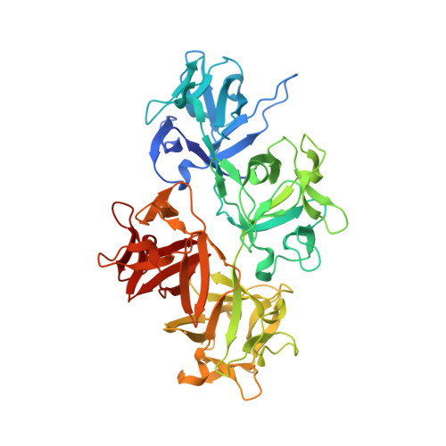
6I18

CRYSTAL STRUCTURE OF FASCIN IN COMPLEX WITH BDP-13176


Experimental Data Snapshot

  • Method: X-RAY DIFFRACTION
  • Resolution: 1.49 Å
  • R-Value Free: 0.195 
  • R-Value Work: 0.172 
  • R-Value Observed: 0.173 

Starting Model: experimental
View more details

wwPDB Validation   3D Report Full Report


Ligand Structure Quality Assessment 


This is version 1.2 of the entry. See complete history


Literature

Structure-based design, synthesis and biological evaluation of a novel series of isoquinolone and pyrazolo[4,3-c]pyridine inhibitors of fascin 1 as potential anti-metastatic agents.

Francis, S.Croft, D.Schuttelkopf, A.W.Parry, C.Pugliese, A.Cameron, K.Claydon, S.Drysdale, M.Gardner, C.Gohlke, A.Goodwin, G.Gray, C.H.Konczal, J.McDonald, L.Mezna, M.Pannifer, A.Paul, N.R.Machesky, L.McKinnon, H.Bower, J.

(2019) Bioorg Med Chem Lett 29: 1023-1029

  • DOI: https://doi.org/10.1016/j.bmcl.2019.01.035
  • Primary Citation of Related Structures:  
    6I0Z, 6I10, 6I11, 6I12, 6I13, 6I14, 6I15, 6I16, 6I17, 6I18

  • PubMed Abstract: 

    Fascin is an actin binding and bundling protein that is not expressed in normal epithelial tissues but overexpressed in a variety of invasive epithelial tumors. It has a critical role in cancer cell metastasis by promoting cell migration and invasion. Here we report the crystal structures of fascin in complex with a series of novel and potent inhibitors. Structure-based elaboration of these compounds enabled the development of a series with nanomolar affinities for fascin, good physicochemical properties and the ability to inhibit fascin-mediated bundling of filamentous actin. These compounds provide promising starting points for fascin-targeted anti-metastatic therapies.


  • Organizational Affiliation

    Drug Discovery Unit, CRUK Beatson Institute, Glasgow G61 1BD, UK. Electronic address: s.francis@beatson.gla.ac.uk.


Macromolecules
Find similar proteins by:  (by identity cutoff)  |  3D Structure
Entity ID: 1
MoleculeChains Sequence LengthOrganismDetailsImage
Fascin493Homo sapiensMutation(s): 0 
Gene Names: FSCN1FAN1HSNSNL
UniProt & NIH Common Fund Data Resources
Find proteins for Q16658 (Homo sapiens)
Explore Q16658 
Go to UniProtKB:  Q16658
PHAROS:  Q16658
GTEx:  ENSG00000075618 
Entity Groups  
Sequence Clusters30% Identity50% Identity70% Identity90% Identity95% Identity100% Identity
UniProt GroupQ16658
Sequence Annotations
Expand
  • Reference Sequence
Small Molecules
Ligands 3 Unique
IDChains Name / Formula / InChI Key2D Diagram3D Interactions
H0N (Subject of Investigation/LOI)
Query on H0N

Download Ideal Coordinates CCD File 
H [auth A]5-[(3,4-dichlorophenyl)methyl]-4-oxidanylidene-1-piperidin-4-yl-~{N}-pyridin-4-yl-pyrazolo[4,3-c]pyridine-7-carboxamide
C24 H22 Cl2 N6 O2
BMOUOZDRMBLNSI-UHFFFAOYSA-N
EDO
Query on EDO

Download Ideal Coordinates CCD File 
C [auth A],
D [auth A],
E [auth A],
F [auth A],
G [auth A]
1,2-ETHANEDIOL
C2 H6 O2
LYCAIKOWRPUZTN-UHFFFAOYSA-N
ACT
Query on ACT

Download Ideal Coordinates CCD File 
B [auth A]ACETATE ION
C2 H3 O2
QTBSBXVTEAMEQO-UHFFFAOYSA-M
Experimental Data & Validation

Experimental Data

  • Method: X-RAY DIFFRACTION
  • Resolution: 1.49 Å
  • R-Value Free: 0.195 
  • R-Value Work: 0.172 
  • R-Value Observed: 0.173 
  • Space Group: P 21 21 21
Unit Cell:
Length ( Å )Angle ( ˚ )
a = 59.66α = 90
b = 87.81β = 90
c = 98.28γ = 90
Software Package:
Software NamePurpose
REFMACrefinement
xia2data scaling
PDB_EXTRACTdata extraction
xia2data reduction

Structure Validation

View Full Validation Report


Ligand Structure Quality Assessment 


Entry History 

Deposition Data

Revision History  (Full details and data files)

  • Version 1.0: 2019-02-27
    Type: Initial release
  • Version 1.1: 2019-03-20
    Changes: Data collection, Database references
  • Version 1.2: 2024-01-24
    Changes: Advisory, Data collection, Database references, Refinement description